Principes physiques de la régénération des vertébrés

Alessandro De Simone

Professeur(e) assistant

  • T: +41 22 379 30 69
  • office 4077b (Sciences III)

Our lab is interested in the physical principles underlying vertebrate regeneration. Regeneration is the astonishing process in which an organism regrows a lost body part. We ask how cells communicate and coordinate with each other for a vertebrate body part to regenerate into its proper form. How are signals organized in time and space and integrated with mechanical forces to orchestrate growth and patterning?

We ask these questions with a quantitative approach at the boundary of physics and biology. We apply experimental, computational, and theoretical methods. We monitor regeneration live, at subcellular resolution and tissue-wide, including in its native context. We use high-sensitivity sensors and reporters to visualize signals, forces and cell dynamics and we analyze data using automated quantification methods. We use theory to conceptualize those complex systems, generate hypotheses and guide the interpretation of experimental data.

Our main model system is zebrafish and we are fond of zebrafish scales. Scales are bone disks that, after loss, regenerate in about two weeks. Regeneration is driven by a monolayer of osteoblasts that regrows and deposits the new bone. We like scales because they are accessible to live imaging, simple enough to be studied quantitatively and have an intriguing regenerative biology. Thus, scales are an ideal system to study how mechano-chemical signals coordinate morphogenesis in regeneration.

We have open positions for Master students, PhD students and postdocs who want to work at the boundary of physics and biology to tackle the mechanisms of regeneration. We are looking for biologists, physicists, engineers, mathematicians, computer scientists and more, interested in developing or strengthening an interdisciplinary profile.


Array of regenerating scales on the trunk of a zebrafish, visualized by the osteoblast marker osx:GFP-CAAX. Dpp: days post-plucking. Scale bar: 1 mm.*

Research projects

Control of scale size and shape by Erk activity waves

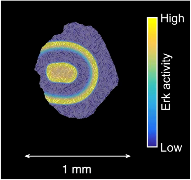
How do signals control the final size of a regenerated tissue? In scale regeneration, cell growth is coordinated by Erk activity waves that originate at the center of the scale and travel across the entire tissue. The more waves are generated, the more the scale will grow. In this project, we ask how waves are generated and propagated, so that scales reach their appropriate final size. What mechanism controls wave generation? Is there a feedback mechanism that informs the waves “source” about tissue size, so that wave generation is tuned accordingly?


Erk activity waves in a regenerating scale. Hpp: hours post-plucking. Scale bar: 250 µm.*

How are signals propagated in a large regenerating tissue? Erk activity waves are compatible with a reaction-diffusion system involving a diffusible Erk activator such as a growth factor, Erk itself and an Erk inhibitor. In this project, we will ask what the molecular components of this mechanism are and how do they interact with each other to propagate waves.


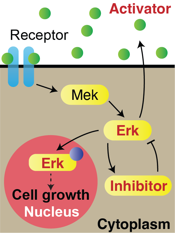
Model of Erk signalling dynamics including a diffusible Erk activator, positive feedback between Erk and the activator, and a negative feedback including an Erk inhibitor. Left: schematics. Right: simulation. Red shaded region: constant source of activator. Scale-bar: 250 µm. *

How does tissue growth lead to form? Erk activity waves impart patterns of tissue growth. Are Erk activity waves a way to distribute growth-induced stresses across the tissues? What are the effects on wave-patterned growth on the final shape of a scale?


Schematics of scale morphogenesis driven by a train of Erk activity waves.*

Coordination of cell growth and bone formation

How are different cell processes coordinated to obtain a regenerated body part that is properly structured? In scale regeneration, osteoblasts deposit bone matrix while they grow, instructed by Erk activity waves. In this project, we will put together the spatial-temporal detail of live imaging with the comprehensive view of transcriptomics to understand how dynamic signals orchestrate different pathways and cell behaviours in regeneration.


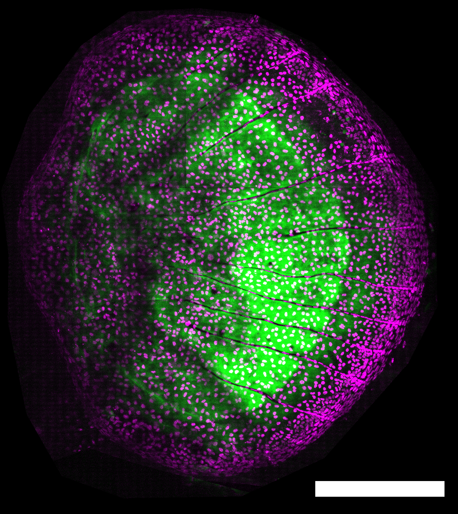
Bone (green) in a regenerating scale (5 days post-plucking; green: Calcein stain; magenta: osx:H2A-mCherry). Scale-bar: 250 µm.

Mechano-chemical coupling in the control of cell proliferation

How is the transition between different phases of regeneration controlled? Put otherwise, how do cells know it is time to change behaviour? During an early phase of scale regeneration, Erk is uniformly active and osteoblasts actively proliferate. Then, Erk switches off in an activation wave that starts at the center of the scale and propagates out. At the same time, osteoblast transition from proliferation to growth without cell division (hypertrophy). In this project, we ask how Erk is patterned over time to control the transition from proliferation to hypertrophy. Our overarching hypothesis is that Erk integrates mechanical inputs from within the tissue and from the neighboring bone. We are relating Erk dynamics, forces, bone biophysical properties and osteoblast behaviour to understand how their interplay controls the final number of cells in a regenerating scale.


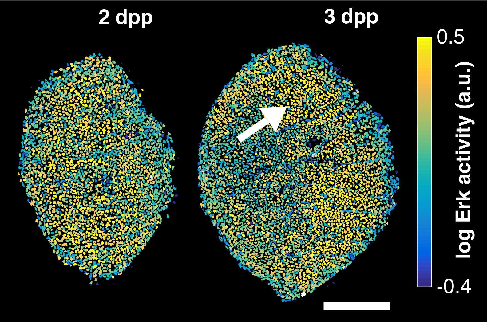
Example of Erk activity in a regenerating scale at 2 and 3 days post-plucking (dpp). Erk activity is activated in a uniform pattern and then switches off starting from the scale centre. Scale-bar: 250 µm. *

*: Figure adapted from De Simone et al., Nature, 2021, DOI: 10.1038/s41586-020-03085-8; Copyright © 2021, The Author(s), under exclusive license to Springer Nature Limited.


External funding

  • SNSF Eccellenza Professorial Fellowship PCEFP3_202776
  • ERC Starting Grant 2021 (funded by SERI)
  • In toto imaging of glial JNK signaling during larval zebrafish spinal cord regeneration.

    Development 2023 Nov;():. 10.1242/dev.202076. 335692.

    résumé

    Identification of signaling events that contribute to innate spinal cord regeneration in zebrafish can inform new targets for modulating injury responses of the mammalian central nervous system. Using a chemical screen, we identify JNK signaling as a necessary regulator of glial cell cycling and tissue bridging during spinal cord regeneration in larval zebrafish. With a kinase translocation reporter, we visualize and quantify JNK signaling dynamics at single-cell resolution in glial cell populations in developing larvae and during injury-induced regeneration. Glial JNK signaling is patterned in time and space during development and regeneration, decreasing globally as the tissue matures and increasing in the rostral cord stump upon transection injury. Thus, dynamic and regional regulation of JNK signaling help to direct glial cell behaviors during innate spinal cord regeneration.

    voir plus de détails sur Pubmed

  • Regeneration: Signaling superhighways.

    Curr Biol 2023 Sep;33(18):R947-R950. 10.1016/j.cub.2023.08.024. S0960-9822(23)01067-9.

    résumé

    Regeneration requires the collective effort of multiple organ systems. A recent study of planarian whole-body regeneration finds that Erk kinase activity propagates rapidly across the entire animal through longitudinal muscle cells to coordinate animal-wide wound responses and that this signal propagation is required for regeneration.

    voir plus de détails sur Pubmed

  • Quantitative Live Imaging of Zebrafish Scale Regeneration: From Adult Fish to Signaling Patterns and Tissue Flows.

    Methods Mol Biol 2024 ;2707():185-204. 10.1007/978-1-0716-3401-1_12.

    résumé

    In regeneration, a damaged body part grows back to its original form. Understanding the mechanisms and physical principles underlying this process has been limited by the difficulties of visualizing cell signals and behaviors in regeneration. Zebrafish scales are emerging as a model system to investigate morphogenesis during vertebrate regeneration using quantitative live imaging. Scales are millimeter-sized dermal bone disks forming a skeletal armor on the body of the fish. The scale bone is deposited by an adjacent monolayer of osteoblasts that, after scale loss, regenerates in about 2 weeks. This intriguing regenerative process is accessible to live confocal microscopy, quantifications, and mathematical modeling. Here, I describe methods to image scale regeneration live, tissue-wide and at sub-cellular resolution. Furthermore, I describe methods to process the resulting images and quantify cell, tissue, and signal dynamics.

    voir plus de détails sur Pubmed

  • Reconstruction and deconstruction of human somitogenesis in vitro.

    Nature 2022 Dec;():. 10.1038/s41586-022-05655-4. 10.1038/s41586-022-05655-4.

    résumé

    The vertebrate body displays a segmental organization which is most conspicuous in the periodic organization of the vertebral column and peripheral nerves. This metameric organization is first implemented when somites, which contain the precursors of skeletal muscles and vertebrae, are rhythmically generated from the presomitic mesoderm (PSM). Somites then become subdivided into anterior and posterior compartments essential for vertebral formation and segmental patterning of the peripheral nervous system. How this key somitic subdivision is established remains poorly understood. Here we introduce novel tridimensional culture systems of human pluripotent stem cells (PSCs), called Somitoids and Segmentoids, which recapitulate the formation of somite-like structures with antero-posterior (AP) identity. We identify a key function of the segmentation clock in converting temporal rhythmicity into the spatial regularity of anterior and posterior somitic compartments. We show that an initial salt-and-pepper expression of the segmentation gene MESP2 in the newly formed segment is transformed into compartments of anterior and posterior identity via an active cell sorting mechanism. Our work demonstrates that the major patterning modules involved in somitogenesis including the clock and wavefront, AP polarity patterning and somite epithelialization can be dissociated and operate independently in our in vitro systems. Together we define a novel framework for the symmetry breaking process initiating somite polarity patterning. Our work provides a valuable platform to decode general principles of somitogenesis and advance knowledge of human development.

    voir plus de détails sur Pubmed

  • Mathematical modeling of Erk activity waves in regenerating zebrafish scales

    Biophys J. 2021 Oct 5;120(19):4287-4297. doi: 10.1016/j.bpj.2021.05.004. Epub 2021 May 20.

    résumé

    Erk signaling regulates cellular decisions in many biological contexts. Recently, we have reported a series of Erk activity traveling waves that coordinate regeneration of osteoblast tissue in zebrafish scales. These waves originate from a central source region, propagate as expanding rings, and impart cell growth, thus controlling tissue morphogenesis. Here, we present a minimal reaction-diffusion model for Erk activity waves. The model considers three components: Erk, a diffusible Erk activator, and an Erk inhibitor. Erk stimulates both its activator and inhibitor, forming a positive and negative feedback loop, respectively. Our model shows that this system can be excitable and propagate Erk activity waves. Waves originate from a pulsatile source that is modeled by adding a localized basal production of the activator, which turns the source region from an excitable to an oscillatory state. As Erk activity periodically rises in the source, it can trigger an excitable wave that travels across the entire tissue. Analysis of the model finds that positive feedback controls the properties of the traveling wavefront and that negative feedback controls the duration of Erk activity peak and the period of Erk activity waves. The geometrical properties of the waves facilitate constraints on the effective diffusivity of the activator, indicating that waves are an efficient mechanism to transfer growth factor signaling rapidly across a large tissue.

    voir plus de détails sur Pubmed

  • Control of osteoblast regeneration by a train of Erk activity waves

    Nature, 590(7844), 129–133. https://doi.org/10.1038/s41586-020-03085-8

    résumé

    Regeneration is a complex chain of events that restores a tissue to its original size and shape. The tissue-wide coordination of cellular dynamics that is needed for proper morphogenesis is challenged by the large dimensions of regenerating body parts. Feedback mechanisms in biochemical pathways can provide effective communication across great distances1-5, but how they might regulate growth during tissue regeneration is unresolved6,7. Here we report that rhythmic travelling waves of Erk activity control the growth of bone in time and space in regenerating zebrafish scales, millimetre-sized discs of protective body armour. We find that waves of Erk activity travel across the osteoblast population as expanding concentric rings that are broadcast from a central source, inducing ring-like patterns of tissue growth. Using a combination of theoretical and experimental analyses, we show that Erk activity propagates as excitable trigger waves that are able to traverse the entire scale in approximately two days and that the frequency of wave generation controls the rate of scale regeneration. Furthermore, the periodic induction of synchronous, tissue-wide activation of Erk in place of travelling waves impairs tissue growth, which indicates that wave-distributed Erk activation is key to regeneration. Our findings reveal trigger waves as a regulatory strategy to coordinate cell behaviour and instruct tissue form during regeneration.

    voir plus de détails sur Pubmed

  • Self-Organized Nuclear Positioning Synchronizes the Cell Cycle in Drosophila Embryos

    Cell. 2019 May 2;177(4):925-941.e17. doi: 10.1016/j.cell.2019.03.007. Epub 2019 Apr 11.

    résumé

    The synchronous cleavage divisions of early embryogenesis require coordination of the cell-cycle oscillator, the dynamics of the cytoskeleton, and the cytoplasm. Yet, it remains unclear how spatially restricted biochemical signals are integrated with physical properties of the embryo to generate collective dynamics. Here, we show that synchronization of the cell cycle in Drosophila embryos requires accurate nuclear positioning, which is regulated by the cell-cycle oscillator through cortical contractility and cytoplasmic flows. We demonstrate that biochemical oscillations are initiated by local Cdk1 inactivation and spread through the activity of phosphatase PP1 to generate cortical myosin II gradients. These gradients cause cortical and cytoplasmic flows that control proper nuclear positioning. Perturbations of PP1 activity and optogenetic manipulations of cortical actomyosin disrupt nuclear spreading, resulting in loss of cell-cycle synchrony. We conclude that mitotic synchrony is established by a self-organized mechanism that integrates the cell-cycle oscillator and embryo mechanics.

    voir plus de détails sur Pubmed

  • In Toto Imaging of Dynamic Osteoblast Behaviors in Regenerating Skeletal Bone

    Curr Biol. 2018 Dec 17;28(24):3937-3947.e4. doi: 10.1016/j.cub.2018.10.052. Epub 2018 Nov 29.

    résumé

    Osteoblasts are matrix-depositing cells that can divide and heal bone injuries. Their deep-tissue location and the slow progression of bone regeneration challenge attempts to capture osteoblast behaviors in live tissue at high spatiotemporal resolution. Here, we have developed an imaging platform to monitor and quantify individual and collective behaviors of osteoblasts in adult zebrafish scales, skeletal body armor discs that regenerate rapidly after loss. Using a panel of transgenic lines that visualize and manipulate osteoblasts, we find that a founder pool of osteoblasts emerges through de novo differentiation within one day of scale plucking. These osteoblasts undergo division events that are largely uniform in frequency and orientation to establish a primordium. Osteoblast proliferation dynamics diversify across the primordium by two days after injury, with cell divisions focused near, and with orientations parallel to, the scale periphery, occurring coincident with dynamic localization of fgf20a gene expression. In posterior scale regions, cell elongation events initiate in areas soon occupied by mineralized grooves called radii, beginning approximately 2 days post injury, with patterned osteoblast death events accompanying maturation of these radii. By imaging at single-cell resolution, we detail acquisition of spatiotemporally distinct cell division, motility, and death dynamics within a founder osteoblast pool as bone regenerates.

    voir plus de détails sur Pubmed

  • Correction: PI(4,5)P forms dynamic cortical structures and directs actin distribution as well as polarity in embryos (doi: 10.1242/dev.164988).

    Development 2018 07;145(13):. 145/13/dev169144. 10.1242/dev.169144.

    résumé

    voir plus de détails sur Pubmed

  • PI(4,5)P 2 forms dynamic cortical structures and directs actin distribution as well as polarity in Caenorhabditis elegans embryos

    Development. 2018 May 30;145(11):dev164988. doi: 10.1242/dev.164988. Erratum in: Development. 2018 Jul 3;145(13).

    résumé

    Asymmetric division is crucial for embryonic development and stem cell lineages. In the one-cell Caenorhabditis elegans embryo, a contractile cortical actomyosin network contributes to asymmetric division by segregating partitioning-defective (PAR) proteins to discrete cortical domains. In the current study, we found that the plasma membrane lipid phosphatidylinositol 4,5-bisphosphate (PIP2) localizes to polarized dynamic structures in C. elegans zygotes, distributing in a PAR-dependent manner along the anterior-posterior (A-P) embryonic axis. PIP2 cortical structures overlap with F-actin, and coincide with the actin regulators RHO-1 and CDC-42, as well as ECT-2. Particle image velocimetry analysis revealed that PIP2 and F-actin cortical movements are coupled, with PIP2 structures moving slightly ahead of F-actin. Importantly, we established that PIP2 cortical structure formation and movement is actin dependent. Moreover, we found that decreasing or increasing the level of PIP2 resulted in severe F-actin disorganization, revealing interdependence between these components. Furthermore, we determined that PIP2 and F-actin regulate the sizing of PAR cortical domains, including during the maintenance phase of polarization. Overall, our work establishes that a lipid membrane component, PIP2, modulates actin organization and cell polarity in C. elegans embryos.

    voir plus de détails sur Pubmed

  • Uncovering the balance of forces driving microtubule aster migration in C. elegans zygotes

    Nat Commun. 2018 Mar 5;9(1):938. doi: 10.1038/s41467-018-03118-x.

    résumé

    Microtubule asters must be positioned precisely within cells. How forces generated by molecular motors such as dynein are integrated in space and time to enable such positioning remains unclear. In particular, whereas aster movements depend on the drag caused by cytoplasm viscosity, in vivo drag measurements are lacking, precluding a thorough understanding of the mechanisms governing aster positioning. Here, we investigate this fundamental question during the migration of asters and pronuclei in C. elegans zygotes, a process essential for the mixing of parental genomes. Detailed quantification of these movements using the female pronucleus as an in vivo probe establish that the drag coefficient of the male-asters complex is approximately five times that of the female pronucleus. Further analysis of embryos lacking cortical dynein, the connection between asters and male pronucleus, or the male pronucleus altogether, uncovers the balance of dynein-driven forces that accurately position microtubule asters in C. elegans zygotes.

    voir plus de détails sur Pubmed

  • Computer simulations reveal mechanisms that organize nuclear dynein forces to separate centrosomes

    Mol Biol Cell. 2017 Nov 7;28(23):3165-3170. doi: 10.1091/mbc.E16-12-0823. Epub 2017 Jul 12.

    résumé

    Centrosome separation along the surface of the nucleus at the onset of mitosis is critical for bipolar spindle assembly. Dynein anchored on the nuclear envelope is known to be important for centrosome separation, but it is unclear how nuclear dynein forces are organized in an anisotropic manner to promote the movement of centrosomes away from each other. Here we use computational simulations of Caenorhabditis elegans embryos to address this fundamental question, testing three potential mechanisms by which nuclear dynein may act. First, our analysis shows that expansion of the nuclear volume per se does not generate nuclear dynein-driven separation forces. Second, we find that steric interactions between microtubules and centrosomes contribute to robust onset of nuclear dynein-mediated centrosome separation. Third, we find that the initial position of centrosomes, between the male pronucleus and cell cortex at the embryo posterior, is a key determinant in organizing microtubule aster asymmetry to power nuclear dynein-dependent separation. Overall our work reveals that accurate initial centrosome position, together with steric interactions, ensures proper anisotropic organization of nuclear dynein forces to separate centrosomes, thus ensuring robust bipolar spindle assembly.

    voir plus de détails sur Pubmed

  • Tension Creates an Endoreplication Wavefront that Leads Regeneration of Epicardial Tissue

    Dev Cell. 2017 Sep 25;42(6):600-615.e4. doi: 10.1016/j.devcel.2017.08.024.

    résumé

    Mechanisms that control cell-cycle dynamics during tissue regeneration require elucidation. Here we find in zebrafish that regeneration of the epicardium, the mesothelial covering of the heart, is mediated by two phenotypically distinct epicardial cell subpopulations. These include a front of large, multinucleate leader cells, trailed by follower cells that divide to produce small, mononucleate daughters. By using live imaging of cell-cycle dynamics, we show that leader cells form by spatiotemporally regulated endoreplication, caused primarily by cytokinesis failure. Leader cells display greater velocities and mechanical tension within the epicardial tissue sheet, and experimentally induced tension anisotropy stimulates ectopic endoreplication. Unbalancing epicardial cell-cycle dynamics with chemical modulators indicated autonomous regenerative capacity in both leader and follower cells, with leaders displaying an enhanced capacity for surface coverage. Our findings provide evidence that mechanical tension can regulate cell-cycle dynamics in regenerating tissue, stratifying the source cell features to improve repair.

    voir plus de détails sur Pubmed

  • Residue-specific Incorporation of Noncanonical Amino Acids into Model Proteins Using an Escherichia coli Cell-free Transcription-translation System.

    J Vis Exp 2016 08;(114):. 10.3791/54273. PMC5091720.

    résumé

    The canonical set of amino acids leads to an exceptionally wide range of protein functionality. Nevertheless, the set of residues still imposes limitations on potential protein applications. The incorporation of noncanonical amino acids can enlarge this scope. There are two complementary approaches for the incorporation of noncanonical amino acids. For site-specific incorporation, in addition to the endogenous canonical translational machineries, an orthogonal aminoacyl-tRNA-synthetase-tRNA pair must be provided that does not interact with the canonical ones. Consequently, a codon that is not assigned to a canonical amino acid, usually a stop codon, is also required. This genetic code expansion enables the incorporation of a noncanonical amino acid at a single, given site within the protein. The here presented work describes residue-specific incorporation where the genetic code is reassigned within the endogenous translational system. The translation machinery accepts the noncanonical amino acid as a surrogate to incorporate it at canonically prescribed locations, i.e., all occurrences of a canonical amino acid in the protein are replaced by the noncanonical one. The incorporation of noncanonical amino acids can change the protein structure, causing considerably modified physical and chemical properties. Noncanonical amino acid analogs often act as cell growth inhibitors for expression hosts since they modify endogenous proteins, limiting in vivo protein production. In vivo incorporation of toxic noncanonical amino acids into proteins remains particularly challenging. Here, a cell-free approach for a complete replacement of L-arginine by the noncanonical amino acid L-canavanine is presented. It circumvents the inherent difficulties of in vivo expression. Additionally, a protocol to prepare target proteins for mass spectral analysis is included. It is shown that L-lysine can be replaced by L-hydroxy-lysine, albeit with lower efficiency. In principle, any noncanonical amino acid analog can be incorporated using the presented method as long as the endogenous in vitro translation system recognizes it.

    voir plus de détails sur Pubmed

  • Dynein Transmits Polarized Actomyosin Cortical Flows to Promote Centrosome Separation

    Cell Rep. 2016 Mar 8;14(9):2250-2262. doi: 10.1016/j.celrep.2016.01.077. Epub 2016 Feb 25.

    résumé

    The two centrosomes present at the onset of mitosis must separate in a timely and accurate fashion to ensure proper bipolar spindle assembly. The minus-end-directed motor dynein plays a pivotal role in centrosome separation, but the underlying mechanisms remain elusive, particularly regarding how dynein coordinates this process in space and time. We addressed these questions in the one-cell C. elegans embryo, using a combination of 3D time-lapse microscopy and computational modeling. Our analysis reveals that centrosome separation is powered by the joint action of dynein at the nuclear envelope and at the cell cortex. Strikingly, we demonstrate that dynein at the cell cortex acts as a force-transmitting device that harnesses polarized actomyosin cortical flows initiated by the centrosomes earlier in the cell cycle. This mechanism elegantly couples cell polarization with centrosome separation, thus ensuring faithful cell division.

    voir plus de détails sur Pubmed

  • Three-dimensional chemotaxis-driven aggregation of tumor cells

    Sci Rep. 2015 Oct 16;5:15205. doi: 10.1038/srep15205.

    résumé

    One of the most important steps in tumor progression involves the transformation from a differentiated epithelial phenotype to an aggressive, highly motile phenotype, where tumor cells invade neighboring tissues. Invasion can occur either by isolated mesenchymal cells or by aggregates that migrate collectively and do not lose completely the epithelial phenotype. Here, we show that, in a three-dimensional cancer cell culture, collective migration of cells eventually leads to aggregation in large clusters. We present quantitative measurements of cluster velocity, coalescence rates, and proliferation rates. These results cannot be explained in terms of random aggregation. Instead, a model of chemotaxis-driven aggregation - mediated by a diffusible attractant - is able to capture several quantitative aspects of our results. Experimental assays of chemotaxis towards culture conditioned media confirm this hypothesis. Theoretical and numerical results further suggest an important role for chemotactic-driven aggregation in spreading and survival of tumor cells.

    voir plus de détails sur Pubmed

  • Cell-free expression with the toxic amino acid canavanine.

    Bioorg Med Chem Lett 2015 Sep;25(17):3658-60. S0960-894X(15)00638-1. 10.1016/j.bmcl.2015.06.045.

    résumé

    Canavanine is a naturally occurring noncanonical amino acid, which is analogous to arginine. It is a potent antimetabolite and natural allelochemic agent, capable of affecting or blocking regulatory and catalytic reactions that involve arginine. Incorporated into proteins at arginine positions, canavanine can be detrimental to protein stability and functional integrity. Although incorporation of canavanine into proteins has long been documented, due to its toxicity, expression in Escherichia coli and other common hosts remains a considerable challenge. Here, we present a simple, cell-free expression system with markedly improved performance compared to heterologous expression. The cell-free expression system does not require any tuning besides substitution of arginine by canavanine. We show that our technique enables highly efficient protein expression in small volumes with arginine being fully replaced by canavanine for functional and structural studies.

    voir plus de détails sur Pubmed

  • Clathrin regulates centrosome positioning by promoting acto-myosin cortical tension in C. elegans embryos

    Development. 2014 Jul;141(13):2712-23. doi: 10.1242/dev.107508.

    résumé

    Regulation of centrosome and spindle positioning is crucial for spatial cell division control. The one-cell Caenorhabditis elegans embryo has proven attractive for dissecting the mechanisms underlying centrosome and spindle positioning in a metazoan organism. Previous work revealed that these processes rely on an evolutionarily conserved force generator complex located at the cell cortex. This complex anchors the motor protein dynein, thus allowing cortical pulling forces to be exerted on astral microtubules emanating from microtubule organizing centers (MTOCs). Here, we report that the clathrin heavy chain CHC-1 negatively regulates pulling forces acting on centrosomes during interphase and on spindle poles during mitosis in one-cell C. elegans embryos. We establish a similar role for the cytokinesis/apoptosis/RNA-binding protein CAR-1 and uncover that CAR-1 is needed to maintain proper levels of CHC-1. We demonstrate that CHC-1 is necessary for normal organization of the cortical acto-myosin network and for full cortical tension. Furthermore, we establish that the centrosome positioning phenotype of embryos depleted of CHC-1 is alleviated by stabilizing the acto-myosin network. Conversely, we demonstrate that slight perturbations of the acto-myosin network in otherwise wild-type embryos results in excess centrosome movements resembling those in chc-1(RNAi) embryos. We developed a 2D computational model to simulate cortical rigidity-dependent pulling forces, which recapitulates the experimental data and further demonstrates that excess centrosome movements are produced at medium cortical rigidity values. Overall, our findings lead us to propose that clathrin plays a critical role in centrosome positioning by promoting acto-myosin cortical tension.

    voir plus de détails sur Pubmed

  • Analysis of precision in chemical oscillators: implications for circadian clocks

    Phys Biol. 2013 Oct;10(5):056005. doi: 10.1088/1478-3975/10/5/056005. Epub 2013 Sep 16.

    résumé

    Biochemical reaction networks often exhibit spontaneous self-sustained oscillations. An example is the circadian oscillator that lies at the heart of daily rhythms in behavior and physiology in most organisms including humans. While the period of these oscillators evolved so that it resonates with the 24 h daily environmental cycles, the precision of the oscillator (quantified via the Q factor) is another relevant property of these cell-autonomous oscillators. Since this quantity can be measured in individual cells, it is of interest to better understand how this property behaves across mathematical models of these oscillators. Current theoretical schemes for computing the Q factors show limitations for both high-dimensional models and in the vicinity of Hopf bifurcations. Here, we derive low-noise approximations that lead to numerically stable schemes also in high-dimensional models. In addition, we generalize normal form reductions that are appropriate near Hopf bifurcations. Applying our approximations to two models of circadian clocks, we show that while the low-noise regime is faithfully recapitulated, increasing the level of noise leads to species-dependent precision. We emphasize that subcomponents of the oscillator gradually decouple from the core oscillator as noise increases, which allows us to identify the subnetworks responsible for robust rhythms.

    voir plus de détails sur Pubmed

Fish Facility Technician position (60-100%) at the University of Geneva

Location: University of Geneva (Switzerland) – Department of Genetics and Evolution, Faculty of Sciences

Closing date: April 15th 2024 (applications will also be considered after that date)

Applications: https://jobs.unige.ch/www/wd_portal.show_job?p_web_site_id=1&p_web_page_id=64939

The De Simone laboratory is looking for a highly motivated animal keeper, to help ensure the smooth running of its animal house, which will house fish and other species in the future.

The animal keeper will be responsible for supervising the breeding, feeding, cleaning feeding, cleaning, organising the health, production and environment of the animals. Seriousness, motivation and punctuality are essential.

You will be responsible for maintaining animal colonies. Your work will include feeding, food preparation, cleaning tanks, monitoring the health and welfare of the animal, water quality monitoring and environmental parameters. Your activities will also include animal breeding, production of transgenic animals, genotyping, biopsies, data and embryo collection, disinfection, participation in experiments and other interventions. You will also be responsible for maintaining the technical equipment organisation of the animal house and its protocols.

The role requires flexibility, excellent organisational and communication skills, and the ability to multi-task.

The position is flexible in terms of hours, as the caretaker will be on call at weekends and during the holidays, and intervene in case of emergencies.

Possession of the CFC, Module 1 Resal/LTK, knowledge of aquatic animals and an excellent knowledge of English would be considerable assets.

The initial contract will be for one year, with the possibility of a longer extension. Salary and benefits are in line with University of Geneva standards.