Laboratoire de régénération et neurogénèse adulte

Brigitte Galliot

Professeur(e) ordinaire

  • T: +41 22 379 67 74
  • office 4055B (Sciences III)

Mots-clés: hydre, cnidaire, régénération, remodelage cellulaire, plasticité du développement, homéostase, blessure, réparation tissulaire, contrôle de la croissance tissulaire, mise en place du schéma corporel, prolifération compensatrice induite par l’apoptose, voies de signalisation, évolution, neurogénèse

1. Résumé

Une question fascinante en biologie est celle posée par la variation du potentiel de régénération à l’intérieur du royaume animal: en effet si de nombreuses espèces sont douées de régénération, la plupart des mammifères ont vraisemblablement perdu ce potentiel. Dans notre laboratoire nous utilisons l’Hydre d’eau douce qui fournit un modèle puissant pour comprendre l’équilibre entre le maintien d’un état homéostatique très dynamique et l’activation de programmes de régénération. Les questions sur lesquelles nous nous penchons sont les suivantes:

  • HOMEOSTASIE et REGENERATION: Quels sont les mécanismes cellulaires et moléculaires qui maintiennent l’homéostasie et quell est leur rôle dans la régénération?
  • CELLULES SOUCHES et CELLULES DIFFERENCIEES: Quelles sont les contributions respectives des cellules souches et des cellules différenciées dans ces processus?
  • MEMOIRE: Quels sont les mécanismes de mémoire qui permettent d’activer le bon programme au bon endroit, c’est à dire la régénération du pied d’un côté, la régénération de la tête de l’autre?
  • NEUROGENESE ADULTE: Quelle est la circuitrie génétique qui conduit à la neurogénèse de novo puisque l’animal reconstitue complètement son système nerveux apical lors de la régénération de la tête?
  • VIEILLISSEMENT: Quels sont les mécanismes de non-vieillissement qui permettent de maintenir accessibles les programmes de développement, régénération et bourgeonnement, quelque soit l’âge de l’animal?
  • EVOLUTION: Lesquels de ces mécanismes ont été conservés au cours de l’évolution?

Afin d’élucider ces questions, nous cherchons à identifier par interférence à l’ARN (ARNi) les cascades de signalisation qui gouvernent la plasticité cellulaire et développementale de l’Hydre. Nous avons récemment montré que la prolifération compensatrice induite par l’apoptose joue un rôle important pour régénérer une tête après bisection au milieu de la colonne corporelle. De façon intéressante, la prolifération compensatrice induite par l’apoptose joue également un rôle important chez le tétard de Xénope qui régénère sa queue, chez la larve de Drosophile qui régénère ses disques imaginaux ou même chez les rongeurs qui régénèrent leur peau ou leur foie. Ces avancées suggèrent qu’il existe des voies communes chez les animaux pour activer un processus de régénération (voir revues récentes [1, 2]).

Figure 1: Schéma montrant la position de l’Hydre dans l’arbre des métazoaires. Parmi les cnidaires, les anthozoaires (coraux, anémonde de mer) sont exclusivement des polypes tandis que les médusozoaires alternant les phases polype et méduse au cours de leur cycle de vie. L’Hydre est un médusozoaire qui a perdu le stade méduse et vit donc exclusivement sous la forme polype.

2. Le modèle de l’Hydre en quelques mots

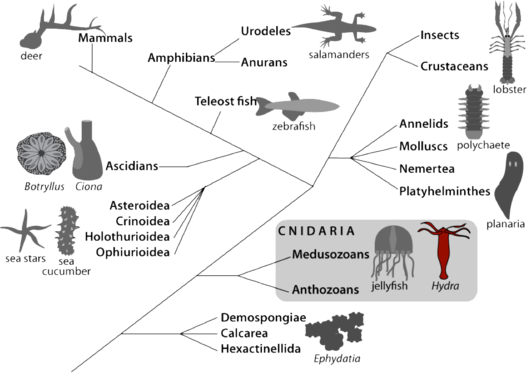
L’Hydre appartient au phylum des cnidaires, un groupe frère des bilatériens (Fig.1). L’anatomie de l’Hydre est simple: c’est en fait un tube qui est différencié aux deux extrémités. Au pôle apical l’animal différencie la région de la tête, formée d’un orifice bouche/anus entouré d’un cercle de tentacules qui servent à attraper la nourriture (l’Hydre est carnivore); au pôle basal, l’animal différencie un disque basal qui lui permet de se fixer. Les tissus sont formés de deux couches cellulaires, l’ectoderme et l’endoderme, séparées par une matrice extracellulaire la mésoglée. L’Hydre differencie tous les types cellulaires nécessaires à la digestion, la sécrétion, la transmission neuro-musculaire, la reproduction sexuée. Ces différents types cellulaires dérivent de trois populations dictinctes de cellules souches: les cellules épithéliales de l’ectoderme, les cellules épithéliales de l’endoderme et les cellules interstitielles qui sont multipotentes [3, 4]. En effet les cellules interstitielles peuvent tout au long de la vie de l’animal se différencier en cellules somatiques (neurones, cellules mécano-sensorielles - cellules urticantes nommées nématocytes ou cnidocytes -, cellules glandulaires) mais aussi en cellules germinales (ovocytes, spermatozoides).

Au cours des 25 dernières années, il a été montré que les gènes codant pour des protéines impliquées dans les voies de signalisation contrôlant les différents comportements cellulaires ou les processus de développement, avaient été extrêmement conservés au cours de l’évolution des métazoaires. Le séquençage du génome de deux espèces de cnidaires (Nematostella vectensis – anémone de mer - et tout récemment Hydra magnipapillata) a confirmé ce haut niveau de conservation génique entre les cnidaires et les vertébrés, renforçant encore la valeur du modèle de l’Hydre [5, 6].

3. Analyse fonctionnelle de la circuitrie génétique soutenant la régénération de l’Hydre

Grâce à l’ARN interférence (ARNi) par nourrissage nous avons pu mettre en évidence des phénotypes partagés par l’homme et l’Hydre

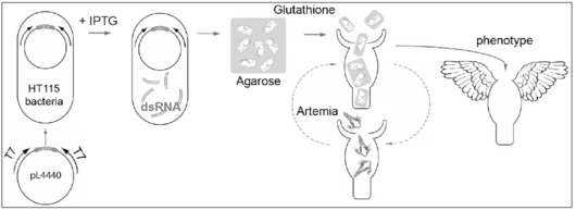
Chez certains vers, il est possible d’éteindre l’expression d’un gène donné en nourrissant les animaux avec des bactéries qui ont au préalable exprimé des ARN double-brins correspondant à ce gène [7-9]. Nous avons adapté cette stratégie à l’Hydre (Fig. 2) et ainsi pu démontrer qu’il s’agit d’une méthode non toxique pour l’Hydre, qui induit progressivement et efficacement des phénotypes spécifiques de chaque gène [10-13].

Dans le cas du gène Kazal1 qui produit un inhibiteur de protéases, l’ARNi induit une autophagie massive des cellules glandulaires qui produisent Kazal1 et des cellules digestives voisines qui normalement utilisent Kazal1. En effet en l’absence de Kazal1, les protéases ne sont pas bloquées et digèrent le contenu des cellules qui les produisent. Les souris mutées pour le gène de type Kazal SPINK3 développent également une autophagie massive au sein de leur pancréas et du tube digestif immédiatement après la naissance. Chez l’homme certaines pancréatites chroniques sont également dues à une autophagie excessive liée à des mutations du gène SPINK1. C’est la preuve de la conservation de la fonction des gènes de type Kazal de l’Hydre à l’homme, soutenant la valeur paradigmatique du système modèle de l’Hydre.

Cette stratégie de l’ARN interférence par nourrissage nous ouvre maintenant la possibilité de cribler un grand nombre de gènes pour identifier leur fonction au cours de la régénération.

Gene silencing by RNA interference in Hydra

Figure 2: Eteignage de l’expression des gènes par ARN interférence par nourrissage chez l’Hydre. Cette stratégie, initialement développée chez le nématode [7, 8] a été adaptée pour les planaires [9] et par notre laboratoire pour les polypes d’Hydre [10-12]. Les ARNs double-brins d’un gène cible sont tout d’abord produits par la bactérie HT115, puis les polypes d’Hydre sont nourris tous les deux jours avec un mélange agarose-bactéries. La glutathione sert à induire l’ouverture de la bouche des polypes. Le nombre de nourrissages requis pour obtenir un phénotype varie suivant le type cellulaire où s’exprime le gène cible, le niveau d’expression de ce gène et la stabilité de la protéine correspondante.

L’Hydre utilise la mort cellulaire et l’activation de la voie Wnt3-bcatenin pour régénérer sa tête

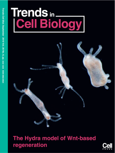
L’activation de la cascade de signalisation Wnt3-bcatenin, qui est parfaitement conservée chez l’Hydre, est nécessaire à la régénération de la tête [14, 15]. Nous avons récemment montré que la bisection induit une vague d’apoptose dans les bourgeons destinés à régénérer une tête mais pas dans ceux destinés à régénérer un pied [13]. Ces cellules en apoptose jouent en fait un rôle moteur dans le processus de régénération car très transitoirement elles délivrent le signal Wnt3 aux cellules adjacentes qui se divisent alors rapidement. En effet si l’apoptose est bloquée ou si la production du signal Wnt3 est inhibée, la prolifération cellulaire et la régénération de la tête sont abolies mais l’addition de signal Wnt3 dans le milieu suffit à sauver le processus de régénération. Une autre expérience a mis en évidence cette fonction de la mort cellulaire dans le processus de régénération de la tête: il suffit d’induire l’apoptose dans les bourgeons destines à régénérer un pied pour activer la voie Wnt3-bcatenin et obtenir des hydres qui régénèrent une tête à la place du pied (Figure 3).

Bi-headed Hydra resulting from the ectopic apoptosis-induced activation of the Wnt3-bcatenin pathway

Figure 3: Hydre à deux têtes résultant de l’induction ectopique de l’apoptose au niveau du bourgeon de régénération du pied. Normalement des Hydres coupées en deux moitiés régénèrent un pied à partir de la moitié apicale et une tête à partir de la moitié basale. Ici des moités apicales ont été brièvement chauffées au niveau de la plaie pour induire un niveau élevé d’apoptose. Cette apoptose ectopique induit alors l’activation de la voie de signalisation Wnt3-bcatenin, ce qui suffit à convertir la régénération du pied en régénération de la tête (voir discussion dans [1, 2]).

La régulation asymmétrique de la cascade MAPK ➜ RSK ➜ CREB ➜ CBP après bisection

La cascade MAPK/CREB semble jouer un rôle précoce dans l’induction de la régénération de la tête. En effet la protéine CREB (cAMP Response Element Binding protein) est un facteur de transcription qui interagit avec des partenaires différents lorsqu’il lie le motif CRE après bisection [16]. De plus CREB est soumis à une série de régulations différentes dans le bourgeon destiné à régénérer une tête et dans celui destine à régénérer un pied: du côté régénérant la tête CREB est immédiatement phosphorylé par la kinase RSK [17] et son expression est rapidement augmentée [18]. Chez les vertébrés, la forme phosphorylée de CREB lie le régulateur chromatinien CBP pour moduler l’expression de gènes cibles. La protéine CBP d’Hydre contient également un domaine liant CREB et lorsque l’expression des gènes RSK, CREB ou CBP est éteinte, ni la vague immédiate d’apoptose, ni le remodelage cellulaire normalement observé dans les bourgeons régénérant une tête n’ont lieu (Chera and Galliot, submitted). Nous cherchons actuellement à comprendre comment cette voie de signalisation permet de “lire” le stress de l’amputation pour activer un programme de régénération spécifique et complexe, celui de la formation de novo de la tête de l’Hydre.

4. L’émergence de la neurogénèse et de la différenciation apicale chez les métazoaires

Chez l’Hydre adulte la neurogénèse est constamment maintenue et le système nerveux apical très dense se forme au cours du bourgeonnement ou bien est complètement régénéré après décapitation [19-21]. Chez les animaux à symétrie bilatérale dont l’ancêtre commun avait déjà un système nerveux centralisé et des organes sensoriels, les gènes à homéoboite des classes ANTP et PAIRED jouent un rôle majeur au cours du développement du système nerveux [22-24]. Chaque classe comprend un grand nombre de familles qui, pour la plupart, se sont diversifiées très tôt au cours de l’évolution animale, précédant l’origine des cnidaires [25-27]. Ceci pose la question de la fonction de ces familles de gènes chez les cnidaires (qui différencient un système nerveux sophistiqué) et chez les porifères (qui n’ont pas de système nerveux) [21].

Les gènes à homéoboite de la classe PAIRED

Parmi les familles de gènes de la classe Paired impliquées dans la spécification du cerveau et des organes sensoriels chez les bilatériens, plusieurs sont exprimées dans le système nerveux des cnidaires [28-32]. Parmi ceux-ci, le gène prd-la est exprimé dans les précurseurs neuronaux et les neurones apicaux de l’Hydre adulte; cependant après bisection, ce gène est précocément et transitoirement exprimé dans la couche endodermale du bourgeon régénérant la tête. Cette double régulation suggère que ce gène est impliqué non seulement dans la neurogénèse mais également dans l’activité organisatrice de la tête en permettant des interactions inductives depuis l’endoderme vers l’ectoderme sus-jacent [19, 28]. Ce résultat est étonnant car chez les vertébrés, certains gènes paired-like ont une fonction similaire lorsque l’activité organisatrice de la tête se met en place au cours du développement précoce, suggérant des mécanismes moléculaires conservés [33].

Les gènes à homéoboite de la classe Hox/ParaHox

Si les cnidaires expriment un grand nombre de familles de gènes de la classe ANTP, toutes les familles Hox utilisées par les bilatéraux ne sont pas présentes et les gènes de type Hox n’ont pas été identifiés chez les porifères, suggérant que ces gènes “Hox-like” de cnidaires représentent en fait les gènes proto-Hox [26, 34-36]. Parmi ceux-ci, le gène cnox-2 (orthologue du gène ParaHox Gsx) est particulièrement intéressant car il est spécifiquement exprimé au cours de la neurogénèse apicale, chez le polype intact mais aussi au cours du bourgeonnement ou de la régénération de la tête [11, 26, 37]. Lorsque l’expression de cnox- 2 est éteinte par ARNi, le système nerveux apical n’est plus renouvellé et ne se forme plus correctement. Chez les vertébrés et chez la mouche Gsx est également impliqué dans la neurogénèse [38]. Ces données suggèrent une fonction neurogénique des gènes cnox- 2/Gsx très conservée au cours de l’évolution ainsi qu’un lien fonctionnel établi très tôt au cours de l’évolution animale entre neurogénèse et différenciation apicale/antérieure [11, 20, 36].

Références

  1. Galliot, B., and Chera, S. (2010) The Hydra model: disclosing an apoptosis-driven generator of Wnt-based regeneration. Trends Cell Biol in press
  2. Galliot, B., and Ghila, L. (2010) Cell plasticity in homeostasis and regeneration. Mol Reprod Dev in press
  3. Steele, R.E. (2002) Developmental signaling in Hydra: what does it take to build a “simple” animal? Dev Biol 248, 199-219
  4. Galliot, B., Miljkovic-Licina, M., de Rosa, R., and Chera, S. (2006) Hydra, a niche for cell and developmental plasticity. Semin Cell Dev Biol 17, 492-502
  5. Putnam, N.H., Srivastava, M., Hellsten, U., Dirks, B., Chapman, J., Salamov, A., Terry, A., Shapiro, H., et al. (2007) Sea anemone genome reveals ancestral eumetazoan gene repertoire and genomic organization. Science 317, 86-94
  6. Chapman, J.A., Kirkness, E.F., Simakov, O., Hampson, S.E., Mitros, T., Weinmaier, T., Rattei, T., Balasubramanian, P.G., et al. (2010) The dynamic genome of Hydra. Nature 464, 592-596
  7. Timmons, L., Court, D.L., and Fire, A. (2001) Ingestion of bacterially expressed dsRNAs can produce specific and potent genetic interference in Caenorhabditis elegans. Gene 263, 103-112
  8. Kamath, R.S., Martinez-Campos, M., Zipperlen, P., Fraser, A.G., and Ahringer, J. (2001) Effectiveness of specific RNA-mediated interference through ingested double-stranded RNA in Caenorhabditis elegans. Genome Biol 2, RESEARCH0002
  9. Newmark, P.A., Reddien, P.W., Cebria, F., and Sanchez Alvarado, A. (2003) Ingestion of bacterially expressed double-stranded RNA inhibits gene expression in planarians. Proc Natl Acad Sci U S A 100 Suppl 1, 11861-11865
  10. Chera, S., de Rosa, R., Miljkovic-Licina, M., Dobretz, K., Ghila, L., Kaloulis, K., and Galliot, B. (2006)
    Silencing of the hydra serine protease inhibitor Kazal1 gene mimics the human SPINK1 pancreatic phenotype. J Cell Sci 119, 846-857
  11. Miljkovic-Licina, M., Chera, S., Ghila, L., and Galliot, B. (2007) Head regeneration in wild-type hydra requires de novo neurogenesis. Development 134, 1191-1201
  12. Buzgariu, W., Chera, S., and Galliot, B. (2008) Methods to investigate autophagy during starvation and regeneration in hydra. Methods Enzymol 451, 409-437
  13. Chera, S., Ghila, L., Dobretz, K., Wenger, Y., Bauer, C., Buzgariu, W., Martinou, J.C., and Galliot, B. (2009) Apoptotic cells provide an unexpected source of Wnt3 signaling to drive hydra head regeneration. Dev Cell 17, 279-289
  14. Hobmayer, B., Rentzsch, F., Kuhn, K., Happel, C.M., von Laue, C.C., Snyder, P., Rothbacher, U., and Holstein, T.W. (2000) WNT signalling molecules act in axis formation in the diploblastic metazoan Hydra. Nature 407, 186-189
  15. Lengfeld, T., Watanabe, H., Simakov, O., Lindgens, D., Gee, L., Law, L., Schmidt, H.A., Ozbek, S., et al. (2009) Multiple Wnts are involved in Hydra organizer formation and regeneration. Dev Biol 330, 186-199
  16. Galliot, B., Welschof, M., Schuckert, O., Hoffmeister, S., and Schaller, H.C. (1995) The cAMP response element binding protein is involved in hydra regeneration. Development 121, 1205-1216
  17. Kaloulis, K., Chera, S., Hassel, M., Gauchat, D., and Galliot, B. (2004) Reactivation of developmental programs: the cAMP-response element-binding protein pathway is involved in hydra head regeneration. Proc Natl Acad Sci U S A 101, 2363-2368
  18. Chera, S., Kaloulis, K., and Galliot, B. (2007) The cAMP response element binding protein (CREB) as an integrative HUB selector in metazoans: clues from the hydra model system. Biosystems 87, 191-203
  19. Miljkovic-Licina, M., Gauchat, D., and Galliot, B. (2004) Neuronal evolution: analysis of regulatory genes in a first-evolved nervous system, the hydra nervous system. Biosystems 76, 75-87
  20. Galliot, B., Quiquand, M., Ghila, L., de Rosa, R., Miljkovic-Licina, M., and Chera, S. (2009) Origins of neurogenesis, a cnidarian view. Dev Biol 332, 2-24
  21. Galliot, B. (2010) A Key Innovation in Evolution, the Emergence of Neurogenesis: Cellular and Molecular Cues from Cnidarian Nervous Systems. In Key Transitions in Animal Evolution (Schierwater, B., and De Salle, R., eds), 127-161, Science Publishers & CRC Press
  22. Pichaud, F., and Desplan, C. (2002) Pax genes and eye organogenesis. Curr Opin Genet Dev 12, 430-434
  23. Hirth, F., Kammermeier, L., Frei, E., Walldorf, U., Noll, M., and Reichert, H. (2003) An urbilaterian origin of the tripartite brain: developmental genetic insights from Drosophila. Development 130, 2365-2373
  24. Denes, A.S., Jekely, G., Steinmetz, P.R., Raible, F., Snyman, H., Prud'homme, B., Ferrier, D.E., Balavoine, G., et al. (2007) Molecular architecture of annelid nerve cord supports common origin of nervous system centralization in bilateria. Cell 129, 277-288
  25. Galliot, B., de Vargas, C., and Miller, D. (1999) Evolution of homeobox genes: Q50 Paired-like genes founded the Paired class. Dev Genes Evol 209, 186-197
  26. Gauchat, D., Mazet, F., Berney, C., Schummer, M., Kreger, S., Pawlowski, J., and Galliot, B. (2000)
    Evolution of Antp-class genes and differential expression of Hydra Hox/paraHox genes in anterior patterning. Proc Natl Acad Sci U S A 97, 4493-4498
  27. Ryan, J.F., Burton, P.M., Mazza, M.E., Kwong, G.K., Mullikin, J.C., and Finnerty, J.R. (2006) The cnidarian-bilaterian ancestor possessed at least 56 homeoboxes. Evidence from the starlet sea anemone, Nematostella vectensis. Genome Biol 7, R64
  28. Gauchat, D., Kreger, S., Holstein, T., and Galliot, B. (1998) prdl-a, a gene marker for hydra apical differentiation related to triploblastic paired-like head-specific genes. Development 125, 1637-1645
  29. Lindgens, D., Holstein, T.W., and Technau, U. (2004) Hyzic, the Hydra homolog of the zic/odd- paired gene, is involved in the early specification of the sensory nematocytes. Development 131, 191-201
  30. Gauchat, D., Escriva, H., Miljkovic-Licina, M., Chera, S., Langlois, M.C., Begue, A., Laudet, V., and Galliot, B. (2004) The orphan COUP-TF nuclear receptors are markers for neurogenesis from cnidarians to vertebrates. Dev Biol 275, 104-123
  31. Stierwald, M., Yanze, N., Bamert, R.P., Kammermeier, L., and Schmid, V. (2004) The Sine oculis/Six class family of homeobox genes in jellyfish with and without eyes: development and eye regeneration. Dev Biol 274, 70-81
  32. Marlow, H.Q., Srivastava, M., Matus, D.Q., Rokhsar, D., and Martindale, M.Q. (2009) Anatomy and development of the nervous system of Nematostella vectensis, an anthozoan cnidarian. Dev Neurobiol 69, 235-254
  33. Galliot, B., and Miller, D. (2000) Origin of anterior patterning. How old is our head? Trends Genet 16, 1-5
  34. Chourrout, D., Delsuc, F., Chourrout, P., Edvardsen, R.B., Rentzsch, F., Renfer, E., Jensen, M.F., Zhu, B., et al. (2006) Minimal ProtoHox cluster inferred from bilaterian and cnidarian Hox complements. Nature 442, 684-687
  35. Chiori, R., Jager, M., Denker, E., Wincker, P., Da Silva, C., Le Guyader, H., Manuel, M., and Queinnec, E. (2009) Are Hox genes ancestrally involved in axial patterning? Evidence from the hydrozoan Clytia hemisphaerica (Cnidaria). PLoS ONE 4, e4231
  36. Quiquand, M., Yanze, N., Schmich, J., Schmid, V., Galliot, B., and Piraino, S. (2009) More constraint on ParaHox than Hox gene families in early metazoan evolution. Dev Biol 328, 173-187
  37. Schummer, M., Scheurlen, I., Schaller, C., and Galliot, B. (1992) HOM/HOX homeobox genes are present in hydra (Chlorohydra viridissima) and are differentially expressed during regeneration. Embo J 11, 1815-1823
  38. Mieko Mizutani, C., and Bier, E. (2008) EvoDevo: the origins of BMP signalling in the neuroectoderm. Nat Rev Genet 9, 663-677
  • The transcription factor Zic4 promotes tentacle formation and prevents epithelial transdifferentiation in Hydra

    Sci Adv 2022 Dec;8(51):eabo0694. 10.1126/sciadv.abo0694.

    résumé

    The molecular mechanisms that maintain cellular identities and prevent dedifferentiation or transdifferentiation remain mysterious. However, both processes are transiently used during animal regeneration. Therefore, organisms that regenerate their organs, appendages, or even their whole body offer a fruitful paradigm to investigate the regulation of cell fate stability. Here, we used as a model system and show that Zic4, whose expression is controlled by Wnt3/β-catenin signaling and the Sp5 transcription factor, plays a key role in tentacle formation and tentacle maintenance. Reducing expression suffices to induce transdifferentiation of tentacle epithelial cells into foot epithelial cells. This switch requires the reentry of tentacle battery cells into the cell cycle without cell division and is accompanied by degeneration of nematocytes embedded in these cells. These results indicate that maintenance of cell fate by a Wnt-controlled mechanism is a key process both during homeostasis and during regeneration.

    voir plus de détails sur Pubmed

  • Combining RNAi-Mediated β-Catenin Inhibition and Reaggregation to Study Hydra Whole-Body Regeneration.

    Methods Mol Biol 2022 ;2450():635-647. 10.1007/978-1-0716-2172-1_34.

    résumé

    In addition to its ability to regenerate any amputated body part, the Hydra freshwater polyp shows the amazing ability to regenerate as a full polyp after a complete dissociation of its tissues. The developmental processes at work in reaggregates undergoing whole-body regeneration can be investigated at the molecular level by RNA interference (RNAi). Here we provide a protocol that combines β-catenin RNAi with reaggregation. This protocol serves as a basis to generate "RNAi-reaggregates," followed by the extraction of high-quality RNA for the precise quantification of gene expression by real-time PCR. This protocol is efficient, providing both a molecular signature, with the significant downregulation of β-catenin and Wnt3, as well as a robust phenotype, the lack of axis formation, which is observed in all reaggregates.

    voir plus de détails sur Pubmed

  • Studying Stem Cell Biology in Intact and Whole-Body Regenerating Hydra by Flow Cytometry.

    Methods Mol Biol 2022 ;2450():373-398. 10.1007/978-1-0716-2172-1_20.

    résumé

    The freshwater Hydra polyp is a versatile model to study whole-body regeneration from a developmental as well as a cellular point of view. The outstanding regenerative capacities of Hydra are based on its three populations of adult stem cells located in the central body column of the animal. There, these three populations, gastrodermal epithelial, epidermal epithelial, and interstitial, continuously cycle in homeostatic conditions, and their activity is locally regulated after mid-gastric bisection. Moreover, they present an unusual cycling behavior with a short G1 phase and a pausing in G2. This particular cell cycle has been studied for a long time with classical microscopic methods. We describe here two flow cytometry methods that provide accurate and reproducible quantitative data to monitor cell cycle regulation in homeostatic and regenerative contexts. We also present a cell sorting procedure based on flow cytometry, whereby stem cells expressing a fluorescent reporter protein in transgenic lines can be enriched for use in applications such as transcriptomic, proteomic, or cell cycle analysis.

    voir plus de détails sur Pubmed

  • Cellular, Metabolic, and Developmental Dimensions of Whole-Body Regeneration in .

    Cold Spring Harb Perspect Biol 2021 Jul;():. cshperspect.a040725. 10.1101/cshperspect.a040725.

    résumé

    Here we discuss the developmental and homeostatic conditions necessary for regeneration. is characterized by populations of adult stem cells paused in the G2 phase of the cell cycle, ready to respond to injury signals. The body column can be compared to a blastema-like structure, populated with multifunctional epithelial stem cells that show low sensitivity to proapoptotic signals, and high inducibility of autophagy that promotes resistance to stress and starvation. Intact polyps also exhibit a dynamic patterning along the oral-aboral axis under the control of homeostatic organizers whose activity results from regulatory loops between activators and inhibitors. As in bilaterians, injury triggers the immediate production of reactive oxygen species (ROS) signals that promote wound healing and contribute to the reactivation of developmental programs via cell death and the de novo formation of new organizing centers from somatic tissues. In aging , regeneration is rapidly lost as homeostatic conditions are no longer pro-regenerative.

    voir plus de détails sur Pubmed

  • The ULK1 kinase, a necessary component of the pro-regenerative and anti-aging machinery in Hydra.

    Mech Ageing Dev 2020 Dec;():111414. S0047-6374(20)30210-4. 10.1016/j.mad.2020.111414.

    résumé

    Hydra vulgaris (Hv) has a high regenerative potential and negligible senescence, as its stem cell populations divide continuously. In contrast, the cold-sensitive H. oligactis (Ho_CS) rapidly develop an aging phenotype under stress, with epithelial stem cells deficient for autophagy, unable to maintain their self-renewal. Here we tested in aging, non-aging and regenerating Hydra the activity and regulation of the ULK1 kinase involved in autophagosome formation. In vitro kinase assays show that human ULK1 activity is activated by Hv extracts but repressed by Ho_CS extracts, reflecting the ability or inability of their respective epithelial cells to initiate autophagosome formation. The factors that keep ULK1 inactive in Ho_CS remain uncharacterized. Hv_Basel1 animals exposed to the ULK1 inhibitor SBI-0206965 no longer regenerate their head, indicating that the sustained autophagy flux recorded in regenerating Hv_AEP2 transgenic animals expressing the DsRed-GFP-LC3A autophagy tandem sensor is necessary. The SBI-0206965 treatment also alters the contractility of intact Hv_Basel1 animals, and leads to a progressive reduction of animal size in Hv_AEP2, similarly to what is observed in ULK1(RNAi) animals. We conclude that the evolutionarily-conserved role of ULK1 in autophagy initiation is crucial to maintain a dynamic homeostasis in Hydra, which supports regeneration efficiency and prevents aging.

    voir plus de détails sur Pubmed

  • The polymorphism of Hydra microsatellite sequences provides strain-specific signatures.

    PLoS ONE 2020 ;15(9):e0230547. 10.1371/journal.pone.0230547. PONE-D-20-06012.

    résumé

    Hydra are freshwater polyps widely studied for their amazing regenerative capacity, adult stem cell populations, low senescence and value as ecotoxicological marker. Many wild-type strains of H. vulgaris have been collected worldwide and maintained effectively under laboratory conditions by asexual reproduction, while stable transgenic lines have been continuously produced since 2006. Efforts are now needed to ensure the genetic characterization of all these strains, which despite similar morphologies, show significant variability in their response to gene expression silencing procedures, pharmacological treatments or environmental conditions. Here, we established a rapid and reliable procedure at the single polyp level to produce via PCR amplification of three distinct microsatellite sequences molecular signatures that distinguish between Hydra strains and species. The TG-rich region of an uncharacterized gene (ms-c25145) helps to distinguish between Eurasian H. vulgaris-Pallas strains (Hm-105, Basel1, Basel2 and reg-16), between Eurasian and North American H. vulgaris strains (H. carnea, AEP), and between the H. vulgaris and H. oligactis species. The AT-rich microsatellite sequences located in the AIP gene (Aryl Hydrocarbon Receptor Interaction Protein, ms-AIP) also differ between Eurasian and North American H. vulgaris strains. Finally, the AT-rich microsatellite located in the Myb-Like cyclin D-binding transcription factor1 gene (ms-DMTF1) gene helps to distinguish certain transgenic AEP lines. This study shows that the analysis of microsatellite sequences, which is capable of tracing genomic variations between closely related lineages of Hydra, provides a sensitive and robust tool for characterizing the Hydra strains.

    voir plus de détails sur Pubmed

  • Deficient autophagy in epithelial stem cells drives aging in the freshwater cnidarian .

    Development 2019 Dec;():. dev.177840. 10.1242/dev.177840.

    résumé

    possesses three distinct stem cell populations that continuously self-renew and prevent aging in However sexual animals from the cold-sensitive ( strain develop an aging phenotype upon gametogenesis induction, initiated by the loss of interstitial stem cells. Animals stop regenerating, lose their active behaviors and die within three months. This phenotype is not observed in the cold-resistant strain. To dissect the mechanisms of aging we compared the self-renewal of epithelial stem cells in these two strains and found it irreversibly reduced in aging while sustained in non-aging We also identified a deficient autophagy in epithelial cells, with a constitutive deficiency in autophagosome formation as detected with the mCherry-eGFP-LC3A/B autophagy sensor, an inefficient response to starvation as evidenced by the accumulation of the autophagosome cargo protein p62/SQSTM1, and a poorly-inducible autophagy flux upon proteasome inhibition. In the non-aging animals, the blockade of autophagy by knocking-down suffices to induce aging. This study highlights the essential role of a dynamic autophagy flux to maintain epithelial stem cell renewal and prevent aging.

    voir plus de détails sur Pubmed

  • Model systems for regeneration: Hydra.

    Development 2019 Nov;146(21):. 146/21/dev177212. 10.1242/dev.177212.

    résumé

    The freshwater polyp provides a potent model system for investigating the conditions that promote wound healing, reactivation of a developmental process and, ultimately, regeneration of an amputated body part. polyps can also be dissociated to the single cell level and can regenerate a complete body axis from aggregates, behaving as natural organoids. In recent years, the ability to exploit has been expanded with the advent of new live-imaging approaches, genetic manipulations that include stable transgenesis, gene silencing and genome editing, and the accumulation of high-throughput omics data. In this Primer, we provide an overview of as a model system for studying regeneration, highlighting recent results that question the classical self-enhancement and long-range inhibition model supposed to drive regeneration. We underscore the need for integrative explanations incorporating biochemical as well as mechanical signalling.

    voir plus de détails sur Pubmed

  • Combining BrdU-Labeling to Detection of Neuronal Markers to Monitor Adult Neurogenesis in Hydra.

    Methods Mol. Biol. 2020 ;2047():3-24. 10.1007/978-1-4939-9732-9_1.

    résumé

    The nervous system is produced and maintained in adult Hydra through the continuous production of nerve cells and mechanosensory cells (nematocytes or cnidocytes). De novo neurogenesis occurs slowly in intact animals that replace their dying nerve cells, at a faster rate in animals regenerating their head as a complete apical nervous system is built in few days. To dissect the molecular mechanisms that underlie these properties, a precise monitoring of the markers of neurogenesis and nematogenesis is required. Here we describe the conditions for an efficient BrdU-labeling coupled to an immunodetection of neuronal markers, either regulators of neurogenesis, here the homeoprotein prdl-a, or neuropeptides such as RFamide or Hym-355. This method can be performed on whole-mount animals as well as on macerated tissues when cells retain their morphology. Moreover, when antibodies are not available, BrdU-labeling can be combined with the analysis of gene expression by whole-mount in situ hybridization. This co-immunodetection procedure is well adapted to visualize and quantify the dynamics of de novo neurogenesis. Upon continuous BrdU labeling, the repeated measurements of BrdU-labeling indexes in specific cellular populations provide a precise monitoring of nematogenesis as well as neurogenesis, in homeostatic or developmental conditions.

    voir plus de détails sur Pubmed

  • Loss of Neurogenesis in Aging Hydra.

    Dev Neurobiol 2019 05;79(5):479-496. 10.1002/dneu.22676. PMC6586502. NIHMS1019840.

    résumé

    In Hydra the nervous system is composed of neurons and mechanosensory cells that differentiate from interstitial stem cells (ISCs), which also provide gland cells and germ cells. The adult nervous system is actively maintained through continuous de novo neurogenesis that occurs at two distinct paces, slow in intact animals and fast in regenerating ones. Surprisingly Hydra vulgaris survive the elimination of cycling interstitial cells and the subsequent loss of neurogenesis if force-fed. By contrast, H. oligactis animals exposed to cold temperature undergo gametogenesis and a concomitant progressive loss of neurogenesis. In the cold-sensitive strain Ho_CS, this loss irreversibly leads to aging and animal death. Within four weeks, Ho_CS animals lose their contractility, feeding response, and reaction to light. Meanwhile, two positive regulators of neurogenesis, the homeoprotein prdl-a and the neuropeptide Hym-355, are no longer expressed, while the "old" RFamide-expressing neurons persist. A comparative transcriptomic analysis performed in cold-sensitive and cold-resistant strains confirms the downregulation of classical neuronal markers during aging but also shows the upregulation of putative regulators of neurotransmission and neurogenesis such as AHR, FGFR, FoxJ3, Fral2, Jagged, Meis1, Notch, Otx1, and TCF15. The switch of Fral2 expression from neurons to germ cells suggests that in aging animals, the neurogenic program active in ISCs is re-routed to germ cells, preventing de novo neurogenesis and impacting animal survival.

    voir plus de détails sur Pubmed

  • An evolutionarily-conserved Wnt3/β-catenin/Sp5 feedback loop restricts head organizer activity in Hydra.

    Nat Commun 2019 01;10(1):312. 10.1038/s41467-018-08242-2. 10.1038/s41467-018-08242-2. PMC6338789.

    résumé

    Polyps of the cnidarian Hydra maintain their adult anatomy through two developmental organizers, the head organizer located apically and the foot organizer basally. The head organizer is made of two antagonistic cross-reacting components, an activator, driving apical differentiation and an inhibitor, preventing ectopic head formation. Here we characterize the head inhibitor by comparing planarian genes down-regulated when β-catenin is silenced to Hydra genes displaying a graded apical-to-basal expression and an up-regulation during head regeneration. We identify Sp5 as a transcription factor that fulfills the head inhibitor properties: leading to a robust multiheaded phenotype when knocked-down in Hydra, acting as a transcriptional repressor of Wnt3 and positively regulated by Wnt/β-catenin signaling. Hydra and zebrafish Sp5 repress Wnt3 promoter activity while Hydra Sp5 also activates its own expression, likely via β-catenin/TCF interaction. This work identifies Sp5 as a potent feedback loop inhibitor of Wnt/β-catenin signaling, a function conserved across eumetazoan evolution.

    voir plus de détails sur Pubmed

  • Modern genomic tools reveal the structural and cellular diversity of cnidarian nervous systems.

    Curr. Opin. Neurobiol. 2019 Jan;56():87-96. S0959-4388(18)30268-X. 10.1016/j.conb.2018.12.004.

    résumé

    Cnidarians shared a common ancestor with bilaterians more than 600 million years ago. This sister group relationship gives them an informative phylogenetic position for understanding the fascinating morphological and molecular cell type diversity of bilaterian nervous systems. Moreover, cnidarians display novel features such as endodermal neurogenesis and independently evolved centralizations, which provide a platform for understanding the evolution of nervous system innovations. In recent years, the application of modern genomic tools has significantly advanced our understanding of cnidarian nervous system structure and function. For example, transgenic reporter lines and gene knockdown experiments in several cnidarian species reveal a significant degree of conservation in the neurogenesis gene regulatory program, while single cell RNA sequencing projects are providing a much deeper understanding of cnidarian neural cell type diversity. At the level of neural function, the physiological properties of ion channels have been described and calcium imaging of the nervous system in whole animals has allowed for the identification of neural circuits underlying specific behaviours. Cnidarians have arrived in the modern era of molecular neurobiology and are primed to provide exciting new insights into the early evolution of nervous systems.

    voir plus de détails sur Pubmed

  • Non-developmental dimensions of adult regeneration in Hydra.

    Int. J. Dev. Biol. 2018 ;62(6-7-8):373-381. 180111bg. 10.1387/ijdb.180111bg.

    résumé

    An essential dimension of 3D regeneration in adult animals is developmental, with the formation of organizers from somatic tissues. These organizers produce signals that recruit surrounding cells and drive the restoration of the missing structures (organs, appendages, body parts). However, even in animals with a high regenerative potential, this developmental potential is not sufficient to achieve regeneration as homeostatic conditions at the time of injury need to be "pro-regenerative". In Hydra, we identified four distinct homeostatic properties that provide a pro-regenerative framework and we discuss here how these non-developmental properties impact regeneration. First, both the epithelial and the interstitial-derived cells are highly plastic along the animal body, a plasticity that offers several routes to achieve regeneration. Second, the abundant stocks of continuously self-renewing adult stem cells form a constitutive pro-blastema in the central body column, readily activated upon bisection. Third, the autophagy machinery in epithelial cells guarantees a high level of fitness and adaptation to detrimental environmental conditions, as evidenced by the loss of regeneration in animals where autophagy is dysfunctional. Fourth, the extracellular matrix, named mesoglea in Hydra, provides a dynamically-patterned environment where the molecular and mechanical signals induced by injury get translated into a regenerative process. We claim that these homeostatic pro-regenerative features contribute to define the high regenerative potential of adult Hydra.

    voir plus de détails sur Pubmed

  • Hydra, a model system for deciphering the mechanisms of aging and resistance to aging.

    Conn’s Handbook for models on human aging (Second Edition); Chapt. 38

    résumé

    Conn's Handbook of Models for Human Aging, Second Edition, presents key aspects of biology, nutrition, factors affecting lifespan, methods of age determination, use in research and the disadvantages/advantages of use. Using a multidisciplinary approach, this updated edition is designed as the only comprehensive, current work that covers the diversity in aging models. Chapters on comparative models explore age-related diseases, including Alzheimer's, joint disease, cataracts, cancer and obesity. Also included are new tricks and approaches not available in primary publications. This must-have handbook is an indispensable resource for researchers interested in the mechanisms of aging, gerontologists, health professionals, allied health practitioners and students.

    voir sur le site externe

  • Impact of cycling cells and cell cycle regulation on Hydra regeneration.

    Dev. Biol. 2018 Jan;433(2):240-253. S0012-1606(17)30413-X. 10.1016/j.ydbio.2017.11.003.

    résumé

    Hydra tissues are made from three distinct populations of stem cells that continuously cycle and pause in G2 instead of G1. To characterize the role of cell proliferation after mid-gastric bisection, we have (i) used flow cytometry and classical markers to monitor cell cycle modulations, (ii) quantified the transcriptomic regulations of 202 genes associated with cell proliferation during head and foot regeneration, and (iii) compared the impact of anti-proliferative treatments on regeneration efficiency. We confirm two previously reported events: an early mitotic wave in head-regenerating tips, when few cell cycle genes are up-regulated, and an early-late wave of proliferation on the second day, preceded by the up-regulation of 17 cell cycle genes. These regulations appear more intense after mid-gastric bisection than after decapitation, suggesting a position-dependent regulation of cell proliferation during head regeneration. Hydroxyurea, which blocks S-phase progression, delays head regeneration when applied before but not after bisection. This result is consistent with the fact that the Hydra central region is enriched in G2-paused adult stem cells, poised to divide upon injury, thus forming a necessary constitutive pro-blastema. However a prolonged exposure to hydroxyurea does not block regeneration as cells can differentiate apical structures without traversing S-phase, and also escape in few days the hydroxyurea-induced S-phase blockade. Thus Hydra head regeneration, which is a fast event, is highly plastic, relying on large stocks of adult stem cells paused in G2 at amputation time, which immediately divide to proliferate and/or differentiate apical structures even when S-phase is blocked.

    voir plus de détails sur Pubmed

  • Trends in tissue repair and regeneration.

    Development 2017 Feb;144(3):357-364. 144/3/357. 10.1242/dev.144279.

    résumé

    The 6th EMBO conference on the Molecular and Cellular Basis of Regeneration and Tissue Repair took place in Paestum (Italy) on the 17th-21st September, 2016. The 160 scientists who attended discussed the importance of cellular and tissue plasticity, biophysical aspects of regeneration, the diverse roles of injury-induced immune responses, strategies to reactivate regeneration in mammals, links between regeneration and ageing, and the impact of non-mammalian models on regenerative medicine.

    voir plus de détails sur Pubmed

  • How Somatic Adult Tissues Develop Organizer Activity.

    Curr. Top. Dev. Biol. 2016 ;116():391-414. S0070-2153(15)00121-0. 10.1016/bs.ctdb.2015.11.002.

    résumé

    The growth and patterning of anatomical structures from specific cellular fields in developing organisms relies on organizing centers that instruct surrounding cells to modify their behavior, namely migration, proliferation, and differentiation. We discuss here how organizers can form in adult organisms, a process of utmost interest for regenerative medicine. Animals like Hydra and planarians, which maintain their shape and fitness thanks to a highly dynamic homeostasis, offer a useful paradigm to study adult organizers in steady-state conditions. Beside the homeostatic context, these model systems also offer the possibility to study how organizers form de novo from somatic adult tissues. Both extracellular matrix remodeling and caspase activation play a key role in this transition, acting as promoters of organizer formation in the vicinity of the wound. Their respective roles and the crosstalk between them just start to be deciphered.

    voir plus de détails sur Pubmed

  • Multi-functionality and plasticity characterize epithelial cells in Hydra.

    Tissue Barriers ;3(4):e1068908. 10.1080/21688370.2015.1068908. 1068908. PMC4681288.

    résumé

    Epithelial sheets, a synapomorphy of all metazoans but porifers, are present as 2 layers in cnidarians, ectoderm and endoderm, joined at their basal side by an extra-cellular matrix named mesoglea. In the Hydra polyp, epithelial cells of the body column are unipotent stem cells that continuously self-renew and concomitantly express their epitheliomuscular features. These multifunctional contractile cells maintain homeostasis by providing a protective physical barrier, by digesting nutrients, by selecting a stable microbiota, and by rapidly closing wounds. In addition, epithelial cells are highly plastic, supporting the adaptation of Hydra to physiological and environmental changes, such as long starvation periods where survival relies on a highly dynamic autophagy flux. Epithelial cells also play key roles in developmental processes as evidenced by the organizer activity they develop to promote budding and regeneration. We propose here an integrative view of the homeostatic and developmental aspects of epithelial plasticity in Hydra.

    voir plus de détails sur Pubmed

  • Loss of neurogenesis in Hydra leads to compensatory regulation of neurogenic and neurotransmission genes in epithelial cells.

    Philos. Trans. R. Soc. Lond., B, Biol. Sci. 2016 Jan;371(1685):20150040. rstb.2015.0040. 10.1098/rstb.2015.0040. PMC4685579.

    résumé

    Hydra continuously differentiates a sophisticated nervous system made of mechanosensory cells (nematocytes) and sensory-motor and ganglionic neurons from interstitial stem cells. However, this dynamic adult neurogenesis is dispensable for morphogenesis. Indeed animals depleted of their interstitial stem cells and interstitial progenitors lose their active behaviours but maintain their developmental fitness, and regenerate and bud when force-fed. To characterize the impact of the loss of neurogenesis in Hydra, we first performed transcriptomic profiling at five positions along the body axis. We found neurogenic genes predominantly expressed along the central body column, which contains stem cells and progenitors, and neurotransmission genes predominantly expressed at the extremities, where the nervous system is dense. Next, we performed transcriptomics on animals depleted of their interstitial cells by hydroxyurea, colchicine or heat-shock treatment. By crossing these results with cell-type-specific transcriptomics, we identified epithelial genes up-regulated upon loss of neurogenesis: transcription factors (Dlx, Dlx1, DMBX1/Manacle, Ets1, Gli3, KLF11, LMX1A, ZNF436, Shox1), epitheliopeptides (Arminins, PW peptide), neurosignalling components (CAMK1D, DDCl2, Inx1), ligand-ion channel receptors (CHRNA1, NaC7), G-Protein Coupled Receptors and FMRFRL. Hence epitheliomuscular cells seemingly enhance their sensing ability when neurogenesis is compromised. This unsuspected plasticity might reflect the extended multifunctionality of epithelial-like cells in early eumetazoan evolution.

    voir plus de détails sur Pubmed

  • The TALE face of Hox proteins in animal evolution.

    Front Genet 2015 ;6():267. 10.3389/fgene.2015.00267. PMC4539518.

    résumé

    Hox genes are major regulators of embryonic development. One of their most conserved functions is to coordinate the formation of specific body structures along the anterior-posterior (AP) axis in Bilateria. This architectural role was at the basis of several morphological innovations across bilaterian evolution. In this review, we traced the origin of the Hox patterning system by considering the partnership with PBC and Meis proteins. PBC and Meis belong to the TALE-class of homeodomain-containing transcription factors and act as generic cofactors of Hox proteins for AP axis patterning in Bilateria. Recent data indicate that Hox proteins acquired the ability to interact with their TALE partners in the last common ancestor of Bilateria and Cnidaria. These interactions relied initially on a short peptide motif called hexapeptide (HX), which is present in Hox and non-Hox protein families. Remarkably, Hox proteins can also recruit the TALE cofactors by using specific PBC Interaction Motifs (SPIMs). We describe how a functional Hox/TALE patterning system emerged in eumetazoans through the acquisition of SPIMs. We anticipate that interaction flexibility could be found in other patterning systems, being at the heart of the astonishing morphological diversity observed in the animal kingdom.

    voir plus de détails sur Pubmed

  • Multifunctionality and plasticity characterize epithelial cells in Hydra

    DOI:10.1080/21688370.2015.1068908

    résumé

    Epithelial sheets, a synapomorphy of all metazoans but porifers, are present as 2 layers in cnidarians, ectoderm and endoderm, joined at their basal side by an extra-cellular matrix named mesoglea. In the Hydra polyp, epithelial cells of the body column are unipotent stem cells that continuously self-renew and concomitantly express their epitheliomuscular features. These multifunctional contractile cells maintain homeostasis by providing a protective physical barrier, by digesting nutrients, by selecting a stable microbiota, and by rapidly closing wounds. In addition, epithelial cells are highly plastic, supporting the adaptation of Hydra to physiological and environmental changes, such as long starvation periods where survival relies on a highly dynamic autophagy flux. Epithelial cells also play key roles in developmental processes as evidenced by the organizer activity they develop to promote budding and regeneration. We propose here an integrative view of the homeostatic and developmental aspects of epithelial plasticity in Hydra.

    voir sur le site externe

  • Hydra, a powerful model for aging studies.

    Invertebr. Reprod. Dev. 2015 Jan;59(sup1):11-16. 10.1080/07924259.2014.927805. 927805. PMC4463768. NIHMS685130.

    résumé

    Cnidarian Hydra polyps escape senescence, most likely due to the robust activity of their three stem cell populations. These stem cells continuously self-renew in the body column and differentiate at the extremities following a tightly coordinated spatial pattern. Paul Brien showed in 1953 that in one particular species, Hydra oligactis, cold-dependent sexual differentiation leads to rapid aging and death. Here, we review the features of this inducible aging phenotype. These cellular alterations, detected several weeks after aging was induced, are characterized by a decreasing density of somatic interstitial cell derivatives, a disorganization of the apical nervous system, and a disorganization of myofibers of the epithelial cells. Consequently, tissue replacement required to maintain homeostasis, feeding behavior, and contractility of the animal are dramatically affected. Interestingly, this aging phenotype is not observed in all H. oligactis strains, thus providing a powerful experimental model for investigations of the genetic control of aging. Given the presence in the cnidarian genome of a large number of human orthologs that have been lost in ecdysozoans, such approaches might help uncover novel regulators of aging in vertebrates.

    voir plus de détails sur Pubmed

  • A dynamic architecture of life.

    F1000Res 2015 ;4():1288. 10.12688/f1000research.7315.1. PMC4760269.

    résumé

    In recent decades, a profound conceptual transformation has occurred comprising different areas of biological research, leading to a novel understanding of life processes as much more dynamic and changeable. Discoveries in plants and animals, as well as novel experimental approaches, have prompted the research community to reconsider established concepts and paradigms. This development was taken as an incentive to organise a workshop in May 2014 at the Academia Nazionale dei Lincei in Rome. There, experts on epigenetics, regeneration, neuroplasticity, and computational biology, using different animal and plant models, presented their insights on important aspects of a dynamic architecture of life, which comprises all organisational levels of the organism. Their work demonstrates that a dynamic nature of life persists during the entire existence of the organism and permits animals and plants not only to fine-tune their response to particular environmental demands during development, but underlies their continuous capacity to do so. Here, a synthesis of the different findings and their relevance for biological thinking is presented.

    voir plus de détails sur Pubmed

  • Injury-induced immune responses in Hydra.

    Semin. Immunol. 2014 Aug;26(4):277-94. S1044-5323(14)00061-X. 10.1016/j.smim.2014.06.004.

    résumé

    The impact of injury-induced immune responses on animal regenerative processes is highly variable, positive or negative depending on the context. This likely reflects the complexity of the innate immune system that behaves as a sentinel in the transition from injury to regeneration. Early-branching invertebrates with high regenerative potential as Hydra provide a unique framework to dissect how injury-induced immune responses impact regeneration. A series of early cellular events likely require an efficient immune response after amputation, as antimicrobial defence, epithelial cell stretching for wound closure, migration of interstitial progenitors toward the wound, cell death, phagocytosis of cell debris, or reconstruction of the extracellular matrix. The analysis of the injury-induced transcriptomic modulations of 2636 genes annotated as immune genes in Hydra identified 43 genes showing an immediate/early pulse regulation in all regenerative contexts examined. These regulations point to an enhanced cytoprotection via ROS signaling (Nrf, C/EBP, p62/SQSMT1-l2), TNFR and TLR signaling (TNFR16-like, TRAF2l, TRAF5l, jun, fos-related, SIK2, ATF1/CREB, LRRC28, LRRC40, LRRK2), proteasomal activity (p62/SQSMT1-l1, Ced6/Gulf, NEDD8-conjugating enzyme Ubc12), stress proteins (CRYAB1, CRYAB2, HSP16.2, DnaJB9, HSP90a1), all potentially regulating NF-κB activity. Other genes encoding immune-annotated proteins such as NPYR4, GTPases, Swap70, the antiproliferative BTG1, enzymes involved in lipid metabolism (5-lipoxygenase, ACSF4), secreted clotting factors, secreted peptidases are also pulse regulated upon bisection. By contrast, metalloproteinases and antimicrobial peptide genes largely follow a context-dependent regulation, whereas the protease inhibitor α2macroglobulin gene exhibits a sustained up-regulation. Hence a complex immune response to injury is linked to wound healing and regeneration in Hydra.

    voir plus de détails sur Pubmed

  • Robust G2 pausing of adult stem cells in Hydra.

    Differentiation ;87(1-2):83-99. S0301-4681(14)00011-5. 10.1016/j.diff.2014.03.001.

    résumé

    Hydra is a freshwater hydrozoan polyp that constantly renews its two tissue layers thanks to three distinct stem cell populations that cannot replace each other, epithelial ectodermal, epithelial endodermal, and multipotent interstitial. These adult stem cells, located in the central body column, exhibit different cycling paces, slow for the epithelial, fast for the interstitial. To monitor the changes in cell cycling in Hydra, we established a fast and efficient flow cytometry procedure, which we validated by confirming previous findings, as the Nocodazole-induced reversible arrest of cell cycling in G2/M, and the mitogenic signal provided by feeding. Then to dissect the cycling and differentiation behaviors of the interstitial stem cells, we used the AEP_cnnos1 and AEP_Icy1 transgenic lines that constitutively express GFP in this lineage. For the epithelial lineages we used the sf-1 strain that rapidly eliminates the fast cycling cells upon heat-shock and progressively becomes epithelial. This study evidences similar cycling patterns for the interstitial and epithelial stem cells, which all alternate between the G2 and S-phases traversing a minimal G1-phase. We also found interstitial progenitors with a shorter G2 that pause in G1/G0. At the animal extremities, most cells no longer cycle, the epithelial cells terminally differentiate in G2 and the interstitial progenitors in G1/G0. At the apical pole ~80% cells are post-mitotic differentiated cells, reflecting the higher density of neurons and nematocytes in this region. We discuss how the robust G2 pausing of stem cells, maintained over weeks of starvation, may contribute to regeneration.

    voir plus de détails sur Pubmed

  • Mechanisms of regeneration. Preface.

    Curr. Top. Dev. Biol. 2014 ;108():xiii-xviii. B978-0-12-391498-9.10000-2. 10.1016/B978-0-12-391498-9.10000-2.

    résumé

    voir plus de détails sur Pubmed

  • Cell death: a program to regenerate.

    Curr. Top. Dev. Biol. 2014 ;108():121-51. B978-0-12-391498-9.00002-4. 10.1016/B978-0-12-391498-9.00002-4.

    résumé

    Recent studies in Drosophila, Hydra, planarians, zebrafish, mice, indicate that cell death can open paths to regeneration in adult animals. Indeed injury can induce cell death, itself triggering regeneration following an immediate instructive mechanism, whereby the dying cells release signals that induce cellular responses over short and/or long-range distances. Cell death can also provoke a sustained derepressing response through the elimination of cells that suppress regeneration in homeostatic conditions. Whether common properties support what we name "regenerative cell death," is currently unclear. As key parameters, we review here the injury proapoptotic signals, the signals released by the dying cells, the cellular responses, and their respective timing. ROS appears as a common signal triggering cell death through MAPK and/or JNK pathway activation. But the modes of ROS production vary, from a brief pulse upon wounding, to repeated waves as observed in the zebrafish fin where ROS supports two peaks of cell death. Indeed regenerative cell death can be restricted to the injury phase, as in Hydra, Drosophila, or biphasic, immediate, and delayed, as in planarians and zebrafish. The dying cells release in a caspase-dependent manner a variety of signaling molecules, cytokines, growth factors, but also prostaglandins or ATP as recorded in Drosophila, Hydra, mice, and zebrafish, respectively. Interestingly, the ROS-producing cells often resist to cell death, implying a complex paracrine mode of signaling to launch regeneration, involving ROS-producing cells, ROS-sensing cells that release signaling molecules upon caspase activation, and effector cells that respond to these signals by proliferating, migrating, and/or differentiating.

    voir plus de détails sur Pubmed

  • Regeneration in Hydra

    eLS, 3rd edition ed. John Wiley & Sons Ltd: Chichester, London. DOI: 10.1002/9780470015902.a0001096.pub3

    résumé

    Hydra freshwater polyps have a remarkable ability to regenerate after bisection or even after dissociation, and thus offer a unique model system to investigate the cellular and molecular basis of eumetazoan regeneration. From a single cut along the body column two different types of regeneration arise: foot regeneration from the apical part and head regeneration from the basal part. The high proportion of stem cells in the Hydra body column supports these fast and efficient processes. Grafting experiments proved that the gastric tissue in the head‐regenerating tip rapidly develops a de novo organising activity, as evidenced by the induction of an ectopic axis when transplanted onto a host. The molecular mechanisms involved in this transformation rely on the immediate activation of the mitogen activated protein kinase (MAPK) pathway and the subsequent activation of the canonical Wnt3 pathway. This early phase is followed by a patterning phase, when head regeneration requires de novo neurogenesis.

    voir sur le site externe

  • Punctuated emergences of genetic and phenotypic innovations in eumetazoan, bilaterian, euteleostome, and hominidae ancestors.

    Genome Biol Evol 2013 ;5(10):1949-68. evt142. 10.1093/gbe/evt142. PMC3814200.

    résumé

    Phenotypic traits derive from the selective recruitment of genetic materials over macroevolutionary times, and protein-coding genes constitute an essential component of these materials. We took advantage of the recent production of genomic scale data from sponges and cnidarians, sister groups from eumetazoans and bilaterians, respectively, to date the emergence of human proteins and to infer the timing of acquisition of novel traits through metazoan evolution. Comparing the proteomes of 23 eukaryotes, we find that 33% human proteins have an ortholog in nonmetazoan species. This premetazoan proteome associates with 43% of all annotated human biological processes. Subsequently, four major waves of innovations can be inferred in the last common ancestors of eumetazoans, bilaterians, euteleostomi (bony vertebrates), and hominidae, largely specific to each epoch, whereas early branching deuterostome and chordate phyla show very few innovations. Interestingly, groups of proteins that act together in their modern human functions often originated concomitantly, although the corresponding human phenotypes frequently emerged later. For example, the three cnidarians Acropora, Nematostella, and Hydra express a highly similar protein inventory, and their protein innovations can be affiliated either to traits shared by all eumetazoans (gut differentiation, neurogenesis); or to bilaterian traits present in only some cnidarians (eyes, striated muscle); or to traits not identified yet in this phylum (mesodermal layer, endocrine glands). The variable correspondence between phenotypes predicted from protein enrichments and observed phenotypes suggests that a parallel mechanism repeatedly produce similar phenotypes, thanks to novel regulatory events that independently tie preexisting conserved genetic modules.

    voir plus de détails sur Pubmed

  • RNAseq versus genome-predicted transcriptomes: a large population of novel transcripts identified in an Illumina-454 Hydra transcriptome.

    BMC Genomics 2013 ;14():204. 1471-2164-14-204. 10.1186/1471-2164-14-204. PMC3764976.

    résumé

    Evolutionary studies benefit from deep sequencing technologies that generate genomic and transcriptomic sequences from a variety of organisms. Genome sequencing and RNAseq have complementary strengths. In this study, we present the assembly of the most complete Hydra transcriptome to date along with a comparative analysis of the specific features of RNAseq and genome-predicted transcriptomes currently available in the freshwater hydrozoan Hydra vulgaris.

    voir plus de détails sur Pubmed

  • Guidelines for the use and interpretation of assays for monitoring autophagy.

    Autophagy 2012 Apr;8(4):445-544. PMC3404883.

    résumé

    In 2008 we published the first set of guidelines for standardizing research in autophagy. Since then, research on this topic has continued to accelerate, and many new scientists have entered the field. Our knowledge base and relevant new technologies have also been expanding. Accordingly, it is important to update these guidelines for monitoring autophagy in different organisms. Various reviews have described the range of assays that have been used for this purpose. Nevertheless, there continues to be confusion regarding acceptable methods to measure autophagy, especially in multicellular eukaryotes. A key point that needs to be emphasized is that there is a difference between measurements that monitor the numbers or volume of autophagic elements (e.g., autophagosomes or autolysosomes) at any stage of the autophagic process vs. those that measure flux through the autophagy pathway (i.e., the complete process); thus, a block in macroautophagy that results in autophagosome accumulation needs to be differentiated from stimuli that result in increased autophagic activity, defined as increased autophagy induction coupled with increased delivery to, and degradation within, lysosomes (in most higher eukaryotes and some protists such as Dictyostelium) or the vacuole (in plants and fungi). In other words, it is especially important that investigators new to the field understand that the appearance of more autophagosomes does not necessarily equate with more autophagy. In fact, in many cases, autophagosomes accumulate because of a block in trafficking to lysosomes without a concomitant change in autophagosome biogenesis, whereas an increase in autolysosomes may reflect a reduction in degradative activity. Here, we present a set of guidelines for the selection and interpretation of methods for use by investigators who aim to examine macroautophagy and related processes, as well as for reviewers who need to provide realistic and reasonable critiques of papers that are focused on these processes. These guidelines are not meant to be a formulaic set of rules, because the appropriate assays depend in part on the question being asked and the system being used. In addition, we emphasize that no individual assay is guaranteed to be the most appropriate one in every situation, and we strongly recommend the use of multiple assays to monitor autophagy. In these guidelines, we consider these various methods of assessing autophagy and what information can, or cannot, be obtained from them. Finally, by discussing the merits and limits of particular autophagy assays, we hope to encourage technical innovation in the field.

    voir plus de détails sur Pubmed

  • Hydra, a fruitful model system for 270 years.

    Int. J. Dev. Biol. 2012 ;56(6-8):411-23. 120086bg. 10.1387/ijdb.120086bg.

    résumé

    The discovery of Hydra regeneration by Abraham Trembley in 1744 promoted much scientific curiosity thanks to his clever design of experimental strategies away from the natural environment. Since then, this little freshwater cnidarian polyp flourished as a potent and fruitful model system. Here, we review some general biological questions that benefitted from Hydra research, such as the nature of embryogenesis, neurogenesis, induction by organizers, sex reversal, symbiosis, aging, feeding behavior, light regulation, multipotency of somatic stem cells, temperature-induced cell death, neuronal transdifferentiation, to cite only a few. To understand how phenotypes arise, theoricists also chose Hydra to model patterning and morphogenetic events, providing helpful concepts such as reaction-diffusion, positional information, and autocatalysis combined with lateral inhibition. Indeed, throughout these past 270 years, scientists used transplantation and grafting experiments, together with tissue, cell and molecular labelings, as well as biochemical procedures, in order to establish the solid foundations of cell and developmental biology. Nowadays, thanks to transgenic, genomic and proteomic tools, Hydra remains a promising model for these fields, but also for addressing novel questions such as evolutionary mechanisms, maintenance of dynamic homeostasis, regulation of stemness, functions of autophagy, cell death, stress response, innate immunity, bioactive compounds in ecosystems, ecotoxicant sensing and science communication.

    voir plus de détails sur Pubmed

  • Preface: the hydra model system.

    Int. J. Dev. Biol. 2012 ;56(6-8):407-9. 120094bg. 10.1387/ijdb.120094bg.

    résumé

    The freshwater Hydra polyp emerged as a model system in 1741 when Abraham Trembley not only discovered its amazing regenerative potential, but also demonstrated that experimental manipulations pave the way to research in biology. Since then, Hydra flourished as a potent and fruitful model system to help answer questions linked to cell and developmental biology, as such as the setting up of an organizer to regenerate a complex missing structure, the establishment and maintainance of polarity in a multicellular organism, the development of mathematical models to explain the robust developmental rules observed in this animal, the maintainance of stemness and multipotency in a highly dynamic environment, the plasticity of differentiated cells, to name but a few. However the Hydra model system is not restricted to cell and developmental biology; during the past 270 years it has also been heavily used to investigate the relationships between Hydra and its environment, opening new horizons concerning neurophysiology, innate immunity, ecosystems, ecotoxicology, symbiosis...

    voir plus de détails sur Pubmed

  • Injury-induced asymmetric cell death as a driving force for head regeneration in Hydra.

    Dev. Genes Evol. 2013 Mar;223(1-2):39-52. 10.1007/s00427-012-0411-y.

    résumé

    The freshwater Hydra polyp provides a unique model system to decipher the mechanisms underlying adult regeneration. Indeed, a single cut initiates two distinct regenerative processes, foot regeneration on one side and head regeneration on the other side, the latter relying on the rapid formation of a local head organizer. Two aspects are discussed here: the asymmetric cellular remodeling induced by mid-gastric bisection and the signaling events that trigger head organizer formation. In head-regenerating tips (but not in foot ones), a wave of cell death takes place immediately, leading the apoptotic cells to transiently release Wnt3 and activate the β-catenin pathway in the neighboring cycling cells to push them through mitosis. This process, which mimics the apoptosis-induced compensatory proliferation process deciphered in Drosophila larvae regenerating their discs, likely corresponds to an evolutionarily conserved mechanism, also at work in Xenopus tadpoles regenerating their tail or mice regenerating their skin or liver. How is this process generated in Hydra? Several studies pointed to the necessary activation of the extracellular signal-regulated kinase (ERK) 1-2 and mitogen-activated protein kinase (MAPK) pathways during early head regeneration. Indeed inhibition of ERK 1-2 or knockdown of RSK, cAMP response element-binding protein (CREB), and CREB-binding protein (CBP) prevent injury-induced apoptosis and head regeneration. The current scenario involves an asymmetric activation of the MAPK/CREB pathway to trigger injury-induced apoptosis in the interstitial cells and in the epithelial cells a CREB/CBP-dependent transcriptional activation of early genes essential for head-organizing activity as wnt3, HyBra1, and prdl-a. The question now is how bisection in the rather uniform central region of the polyp can generate this immediately asymmetric signaling.

    voir plus de détails sur Pubmed

  • How to use Hydra as a model system to teach biology in the classroom.

    Int. J. Dev. Biol. 2012 ;56(6-8):637-52. 123523pb. 10.1387/ijdb.123523pb.

    résumé

    As scientists it is our duty to fight against obscurantism and loss of rational thinking if we want politicians and citizens to freely make the most intelligent choices for the future generations. With that aim, the scientific education and training of young students is an obvious and urgent necessity. We claim here that Hydra provides a highly versatile but cheap model organism to study biology at any age. Teachers of biology have the unenviable task of motivating young people, who with many other motivations that are quite valid, nevertheless must be guided along a path congruent with a 'syllabus' or a 'curriculum'. The biology of Hydra spans the history of biology as an experimental science from Trembley's first manipulations designed to determine if the green polyp he found was plant or animal to the dissection of the molecular cascades underpinning, regeneration, wound healing, stemness, aging and cancer. It is described here in terms designed to elicit its wider use in classrooms. Simple lessons are outlined in sufficient detail for beginners to enter the world of 'Hydra biology'. Protocols start with the simplest observations to experiments that have been pretested with students in the USA and in Europe. The lessons are practical and can be used to bring 'life', but also rational thinking into the study of life for the teachers of students from elementary school through early university.

    voir plus de détails sur Pubmed

  • Hydra, a versatile model to study the homeostatic and developmental functions of cell death.

    Int. J. Dev. Biol. 2012 ;56(6-8):593-604. 123499sr. 10.1387/ijdb.123499sr.

    résumé

    In the freshwater cnidarian polyp Hydra, cell death takes place in multiple contexts. Indeed apoptosis occurs during oogenesis and spermatogenesis, during starvation, and in early head regenerating tips, promoting local compensatory proliferation at the boundary between heterografts. Apoptosis can also be induced upon exposure to pro-apoptotic agents (colchicine, wortmannin), upon heat-shock in the thermosensitive sf-1 mutant, and upon wounding. In all these contexts, the cells that undergo cell death belong predominantly to the interstitial cell lineage, whereas the epithelial cells, which are rather resistant to pro-apoptotic signals, engulf the apoptotic bodies. Beside this clear difference between the interstitial and the epithelial cell lineages, the different interstitial cell derivatives also show noticeable variations in their respective apoptotic sensitivity, with the precursor cells appearing as the most sensitive to pro-apoptotic signals. The apoptotic machinery has been well conserved across evolution. However, its specific role and regulation in each context are not known yet. Tools that help characterize apoptotic activity in Hydra have recently been developed. Among them, the aposensor Apoliner initially designed in Drosophila reliably measures wortmannin-induced apoptotic activity in a biochemical assay. Also, flow cytometry and TUNEL analyses help identify distinctive features between wortmannin-induced and heat-shock induced apoptosis in the sf-1 strain. Thanks to the live imaging tools already available, Hydra now offers a model system with which the functions of the apoptotic machinery to maintain long-term homeostasis, stem cell renewal, germ cell production, active developmental processes and non-self response can be deciphered.

    voir plus de détails sur Pubmed

  • A two-step process in the emergence of neurogenesis.

    Eur. J. Neurosci. 2011 Sep;34(6):847-62. 10.1111/j.1460-9568.2011.07829.x.

    résumé

    Cnidarians belong to the first phylum differentiating a nervous system, thus providing suitable model systems to trace the origins of neurogenesis. Indeed corals, sea anemones, jellyfish and hydra contract, swim and catch their food thanks to sophisticated nervous systems that share with bilaterians common neurophysiological mechanisms. However, cnidarian neuroanatomies are quite diverse, and reconstructing the urcnidarian nervous system is ambiguous. At least a series of characters recognized in all classes appear plesiomorphic: (1) the three cell types that build cnidarian nervous systems (sensory-motor cells, ganglionic neurons and mechanosensory cells called nematocytes or cnidocytes); (2) an organization of nerve nets and nerve rings [those working as annular central nervous system (CNS)]; (3) a neuronal conduction via neurotransmitters; (4) a larval anterior sensory organ required for metamorphosis; (5) a persisting neurogenesis in adulthood. By contrast, the origin of the larval and adult neural stem cells differs between hydrozoans and other cnidarians; the sensory organs (ocelli, lens-eyes, statocysts) are present in medusae but absent in anthozoans; the electrical neuroid conduction is restricted to hydrozoans. Evo-devo approaches might help reconstruct the neurogenic status of the last common cnidarian ancestor. In fact, recent genomic analyses show that if most components of the postsynaptic density predate metazoan origin, the bilaterian neurogenic gene families originated later, in basal metazoans or as eumetazoan novelties. Striking examples are the ParaHox Gsx, Pax, Six, COUP-TF and Twist-type regulators, which seemingly exert neurogenic functions in cnidarians, including eye differentiation, and support the view of a two-step process in the emergence of neurogenesis.

    voir plus de détails sur Pubmed

  • [Between homeostasis and development, which strategies to regenerate?].

    Biol Aujourdhui 2011 ;205(2):125-37. 10.1051/jbio/2011011. jbio2011011.

    résumé

    The Hydra model system is well suited to decipher the mechanisms underlying adult regeneration, specifically those that were robust enough to be maintained across evolution. After mid-gastric bissection head regeneration in Hydra relies on apoptosis-induced compensatory proliferation via the release of Wnt3 by the apoptotic interstitial cells and activation of the β-catenin pathway in the surrounding cycling interstitial cells. As apoptosis-induced compensatory proliferation is also at work in Drosophila regenerating imaginal discs, Xenopus tadpole regenerating their tail and mice regenerating their skin or their liver, this mechanism might represent an evolutionarily-conserved way to launch a regenerative response. However after decapitation, the analysis of the activation of the canonical Wnt pathway in decapitated Hydra showed that apoptosis-induced compensatory proliferation does not take place in this context. Given that the proportion of interstitial stem cells is significantly higher in the middle part than in the upper part of the body column, this suggested that the route taken to regenerate a structure as complex as the head dramatically varies according to the homeostatic status of the tissue at the time of injury. From these observations, we propose a tri-modular model for animal regeneration where the first module or "wound healing module" is followed by a transient module named "inducing module of regeneration" that allows the recruitment of the third module named "re-development module", necessary for repatterning the missing structure. We claim that among these three modules, the inducing module of regeneration is the most drastically constrained by the homeostatic conditions of any given tissue or organ at the time of injury and therefore the most variable.

    voir plus de détails sur Pubmed

  • Injury-induced activation of the MAPK/CREB pathway triggers apoptosis-induced compensatory proliferation in hydra head regeneration.

    Dev. Growth Differ. 2011 Feb;53(2):186-201. 10.1111/j.1440-169X.2011.01250.x.

    résumé

    After bisection, Hydra polyps regenerate their head from the lower half thanks to a head-organizer activity that is rapidly established at the tip. Head regeneration is also highly plastic as both the wild-type and the epithelial Hydra (that lack the interstitial cell lineage) can regenerate their head. In the wild-type context, we previously showed that after mid-gastric bisection, a large subset of the interstitial cells undergo apoptosis, inducing compensatory proliferation of the surrounding progenitors. This asymmetric process is necessary and sufficient to launch head regeneration. The apoptotic cells transiently release Wnt3, which promotes the formation of a proliferative zone by activating the beta-catenin pathway in the adjacent cycling cells. However the injury-induced signaling that triggers apoptosis is unknown. We previously reported an asymmetric immediate activation of the mitogen-activated protein kinase/ribosomal S6 kinase/cAMP response element binding protein (MAPK/RSK/CREB) pathway in head-regenerating tips after mid-gastric bisection. We show here that pharmacological inhibition of the MAPK/ERK pathway or RNAi knockdown of the RSK, CREB, CREB binding protein (CBP) genes prevents apoptosis, compensatory proliferation and blocks head regeneration. As the activation of the MAPK pathway upon injury plays an essential role in regenerating bilaterian species, these results suggest that the MAPK-dependent activation of apoptosis-induced compensatory proliferation represents an evolutionary-conserved mechanism to launch a regenerative process.

    voir plus de détails sur Pubmed

  • Information processing in cells and tissues (IPCAT'2009): from small scale dynamics to understanding systems behavior.

    BioSystems 2010 Oct;102(1):1-2. S0303-2647(10)00123-1. 10.1016/j.biosystems.2010.07.015.

    résumé

    voir plus de détails sur Pubmed

  • The Hydra model: disclosing an apoptosis-driven generator of Wnt-based regeneration.

    Trends Cell Biol. 2010 Sep;20(9):514-23. S0962-8924(10)00099-1. 10.1016/j.tcb.2010.05.006.

    résumé

    The Hydra model system is well suited for the eludication of the mechanisms underlying regeneration in the adult, and an understanding of the core mechanisms is likely to cast light on pathways conserved in other species. Recent detailed analyses of the activation of the Wnt-beta-catenin pathway in bisected Hydra shows that the route taken to regenerate a structure as complex as the head varies dramatically according to the level of the amputation. When decapitation induces direct re-development due to Wnt3 signaling from epithelial cells, head regeneration after mid-gastric section relies first on Wnt3 signaling from interstitial cells, that undergo apoptosis-induced compensatory proliferation, and subsequently on activation of Wnt3 signaling in the epithelial cells. The relative distribution between stem cells and head progenitor cells is strikingly different in these two contexts, indicating that the pre-amputation homeostatic conditions define and constrain the route that bridges wound-healing to the re-development program of the missing structure.

    voir plus de détails sur Pubmed

  • Cell plasticity in homeostasis and regeneration.

    Mol. Reprod. Dev. 2010 Oct;77(10):837-55. 10.1002/mrd.21206.

    résumé

    Over the past decades, genetic analyses performed in vertebrate and invertebrate organisms deciphered numerous cellular and molecular mechanisms deployed during sexual development and identified genetic circuitries largely shared among bilaterians. In contrast, the functional analysis of the mechanisms that support regenerative processes in species randomly scattered among the animal kingdom, were limited by the lack of genetic tools. Consequently, unifying principles explaining how stress and injury can lead to the reactivation of a complete developmental program with restoration of original shape and function remained beyond reach of understanding. Recent data on cell plasticity suggest that beside the classical developmental approach, the analysis of homeostasis and asexual reproduction in adult organisms provides novel entry points to dissect the regenerative potential of a given species, a given organ or a given tissue. As a clue, both tissue homeostasis and regeneration dynamics rely on the availability of stem cells and/or on the plasticity of differentiated cells to replenish the missing structure. The freshwater Hydra polyp provides us with a unique model system to study the intricate relationships between the mechanisms that regulate the maintenance of homeostasis, even in extreme conditions (starvation and overfeeding) and the reactivation of developmental programs after bisection or during budding. Interestingly head regeneration in Hydra can follow several routes according to the level of amputation, suggesting that indeed the homeostatic background dramatically influences the route taken to bridge injury and regeneration.

    voir plus de détails sur Pubmed

  • A Key Innovation in Animal Evolution, the Emergence of Neurogenesis: Cellular and Molecular Cues from Cnidarian Nervous Systems

    BOOK CHAPTER: In Key Transitions in Animal Evolution, Edited by B. Schierwater, and R. De Salle. ISBN 978-1-57808-695-5

    résumé

    The emergence of neurogenesis led to the acquisition of an efficient neuromuscular transmission in eumetazoans, as shown by cnidarians that use evolutionarily-conserved neurophysiological principles to crumple, feed, swim. However, the cnidarian neuroanatomies are quite diverse and reconstructing the urcnidarian nervous system is not an easy task. Three types of characters shared by anthozoans and medusozoans appear plesiomorphic: 1) three cell types that all cnidarians differentiate, neurosensory cells, ganglionic neurons and nematocytes (cnidocytes) that combine mechano-chemosensation and venom secretion; 2) a chemical conduction through nerve nets and nerve rings, those being considered as annular central nervous systems; 3) a larval apical sensory organ that initiates metamorphosis. Other characters receive a disputed origin: 1) the neural stem cell(s), multipotent interstitial stem cell in hydrozoans, not identified in other classes; 2) the electrical conduction through neurons and epithelial cells present only in hydrozoans; 3) the embryonic origin of the nervous system; 4) the medusa sensory organs, ocelli or lens-eyes for light, statocysts for pressure, lacking in anthozoans. Nevertheless numerous gene families that regulate bilaterian neurogenesis are expressed during cnidarian neurogenesis, e.g. cnidarian eyes express Pax, Six and opsin, supporting a common origin for vision. However data establishing a clear picture of the cnidarian neurogenic circuitry are currently missing. Finally many “neurogenic” gene families likely arose and evolved in the absence of neurogenesis, as exemplified by Porifera that express them but lack synaptic transmission. Therefore some eumetazoan-specific families, missing in Porifera as ParaHox/Hox-like and Otx-like genes, might have contributed to the emergence of neurogenesis.

    voir sur le site externe

  • Apoptotic cells provide an unexpected source of Wnt3 signaling to drive hydra head regeneration.

    Dev. Cell 2009 Aug;17(2):279-89. S1534-5807(09)00298-6. 10.1016/j.devcel.2009.07.014.

    résumé

    Decapitated Hydra regenerate their heads via morphallaxis, i.e., without significant contributions made by cell proliferation or interstitial stem cells. Indeed, Hydra depleted of interstitial stem cells regenerate robustly, and Wnt3 from epithelial cells triggers head regeneration. However, we find a different mechanism controlling regeneration after midgastric bisection in animals equipped with both epithelial and interstitial cell lineages. In this context, we see rapid induction of apoptosis and Wnt3 secretion among interstitial cells at the head- (but not foot-) regenerating site. Apoptosis is both necessary and sufficient to induce Wnt3 production and head regeneration, even at ectopic sites. Further, we identify a zone of proliferation beneath the apoptotic zone, reminiscent of proliferative blastemas in regenerating limbs and of compensatory proliferation induced by dying cells in Drosophila imaginal discs. We propose that different types of injuries induce distinct cellular modes of Hydra head regeneration, which nonetheless converge on a central effector, Wnt3.

    voir plus de détails sur Pubmed

  • Origins of neurogenesis, a cnidarian view.

    Dev. Biol. 2009 Aug;332(1):2-24. S0012-1606(09)00875-6. 10.1016/j.ydbio.2009.05.563.

    résumé

    New perspectives on the origin of neurogenesis emerged with the identification of genes encoding post-synaptic proteins as well as many "neurogenic" regulators as the NK, Six, Pax, bHLH proteins in the Demosponge genome, a species that might differentiate sensory cells but no neurons. However, poriferans seem to miss some key regulators of the neurogenic circuitry as the Hox/paraHox and Otx-like gene families. Moreover as a general feature, many gene families encoding evolutionarily-conserved signaling proteins and transcription factors were submitted to a wave of gene duplication in the last common eumetazoan ancestor, after Porifera divergence. In contrast gene duplications in the last common bilaterian ancestor, Urbilateria, are limited, except for the bHLH Atonal-class. Hence Cnidaria share with Bilateria a large number of genetic tools. The expression and functional analyses currently available suggest a neurogenic function for numerous orthologs in developing or adult cnidarians where neurogenesis takes place continuously. As an example, in the Hydra polyp, the Clytia medusa and the Acropora coral, the Gsx/cnox2/Anthox-2 ParaHox gene likely supports neurogenesis. Also neurons and nematocytes (mechanosensory cells) share in hydrozoans a common stem cell and several regulatory genes indicating that they can be considered as sister cells. Performed in anthozoan and medusozoan species, these studies should tell us more about the way(s) evolution hazards achieved the transition from epithelial to neuronal cell fate, and about the robustness of the genetic circuitry that allowed neuromuscular transmission to arise and be maintained across evolution.

    voir plus de détails sur Pubmed

  • More constraint on ParaHox than Hox gene families in early metazoan evolution.

    Dev. Biol. 2009 Apr;328(2):173-87. S0012-1606(09)00062-1. 10.1016/j.ydbio.2009.01.022.

    résumé

    Hox and ParaHox (H/P) genes belong to evolutionary-sister clusters that arose through duplication of a ProtoHOX cluster early in animal evolution. In contrast to bilaterians, cnidarians express, beside PG1, PG2 and Gsx orthologs, numerous Hox-related genes with unclear origin. We characterized from marine hydrozoans three novel Hox-related genes expressed at medusa and polyp stages, which include a Pdx/Xlox ParaHox ortholog induced 1 day later than Gsx during embryonic development. To reconstruct H/P genes' early evolution, we performed multiple systematic comparative phylogenetic analyses, which identified derived sequences that blur the phylogenetic picture, recorded dramatically different evolutionary rates between ParaHox and Hox in cnidarians and showed the unexpected grouping of [Gsx-Pdx/Xlox-PG2-PG3] families in a single metagroup distinct from PG1. We propose a novel more parsimonious evolutionary scenario whereby H/P genes originated from a [Gsx-Pdx/Xlox-PG2-PG3]-related ProtoHox gene, the "posterior" and "anterior" H/P genes appearing secondarily. The ProtoHOX cluster would have contained the three Gsx/PG2, Pdx/PG3, Cdx/PG9 paralogs and produced through tandem duplication the primordial HOX and ParaHOX clusters in the Cnidaria-Bilateria ancestor. The stronger constraint on cnidarian ParaHox genes suggests that the primary function of pre-bilaterian H/P genes was to drive cellular evolutionary novelties such as neurogenesis rather than axis specification.

    voir plus de détails sur Pubmed

  • Autophagy in Hydra: a response to starvation and stress in early animal evolution.

    Biochim. Biophys. Acta 2009 Sep;1793(9):1432-43. S0167-4889(09)00082-2. 10.1016/j.bbamcr.2009.03.010.

    résumé

    The Hydra polyp provides a powerful model system to investigate the regulation of cell survival and cell death in homeostasis and regeneration as Hydra survive weeks without feeding and regenerates any missing part after bisection. Induction of autophagy during starvation is the main surviving strategy in Hydra as autophagic vacuoles form in most myoepithelial cells after several days. When the autophagic process is inhibited, animal survival is actually rapidly jeopardized. An appropriate regulation of autophagy is also essential during regeneration as Hydra RNAi knocked-down for the serine protease inhibitor Kazal-type (SPINK) gene Kazal1, exhibit a massive autophagy after amputation that rapidly compromises cell and animal survival. This excessive autophagy phenotype actually mimics that observed in the mammalian pancreas when SPINK genes are mutated, highlighting the paradigmatic value of the Hydra model system for deciphering pathological processes. Interestingly autophagy during starvation predominantly affects ectodermal epithelial cells and lead to cell survival whereas Kazal1(RNAi)-induced autophagy is restricted to endodermal digestive cells that rapidly undergo cell death. This indicates that distinct regulations that remain to be identified, are at work in these two contexts. Cnidarian express orthologs for most components of the autophagy and TOR pathways suggesting evolutionarily-conserved roles during starvation.

    voir plus de détails sur Pubmed

  • Methods to investigate autophagy during starvation and regeneration in hydra.

    Meth. Enzymol. 2008 ;451():409-37. S0076-6879(08)03226-6. 10.1016/S0076-6879(08)03226-6.

    résumé

    In hydra, the regulation of the balance between cell death and cell survival is essential to maintain homeostasis across the animal and promote animal survival during starvation. Moreover, this balance also appears to play a key role during regeneration of the apical head region. The recent finding that autophagy is a crucial component of this balance strengthens the value of the Hydra model system to analyze the implications of autophagy in starvation, stress response and regeneration. We describe here how we adapted to Hydra some established tools to monitor steady-state autophagy. The ATG8/LC3 marker used in biochemical and immunohistochemical analyses showed a significant increase in autophagosome formation in digestive cells after 11 days of starvation. Moreover, the maceration procedure that keeps intact the morphology of the various cell types allows the quantification of the autophagosomes and autolysosomes in any cell type, thanks to the detection of the MitoFluor or LysoTracker dyes combined with the anti-LC3, anti-LBPA, and/or anti-RSK (ribosomal S6 kinase) immunostaining. The classical activator (rapamycin) and inhibitors (wortmannin, bafilomycin A(1)) of autophagy also appear to be valuable tools to modulate autophagy in hydra, as daily-fed and starved hydra display slightly different responses. Finally, we show that the genetic circuitry underlying autophagy can be qualitatively and quantitatively tested through RNA interference in hydra repeatedly exposed to double-stranded RNAs.

    voir plus de détails sur Pubmed

  • Triggering the regeneration and tissue repair programs.

    Development 2009 Feb;136(3):349-53. 136/3/349. 10.1242/dev.031682.

    résumé

    In early October 2008, researchers from diverse backgrounds gathered at an EMBO conference entitled 'The Molecular and Cellular Basis of Regeneration and Tissue Repair' to discuss the basic biology of regeneration. Topics included cell plasticity in regenerative and developmental contexts, and the link between wound healing and regeneration. The meeting also highlighted the progress made in identifying the molecular networks that underlie regeneration in a variety of model systems.

    voir plus de détails sur Pubmed

  • Guidelines for the use and interpretation of assays for monitoring autophagy in higher eukaryotes.

    Autophagy 2008 Feb;4(2):151-75. 5338. PMC2654259. NIHMS96070.

    résumé

    Research in autophagy continues to accelerate,(1) and as a result many new scientists are entering the field. Accordingly, it is important to establish a standard set of criteria for monitoring macroautophagy in different organisms. Recent reviews have described the range of assays that have been used for this purpose.(2,3) There are many useful and convenient methods that can be used to monitor macroautophagy in yeast, but relatively few in other model systems, and there is much confusion regarding acceptable methods to measure macroautophagy in higher eukaryotes. A key point that needs to be emphasized is that there is a difference between measurements that monitor the numbers of autophagosomes versus those that measure flux through the autophagy pathway; thus, a block in macroautophagy that results in autophagosome accumulation needs to be differentiated from fully functional autophagy that includes delivery to, and degradation within, lysosomes (in most higher eukaryotes) or the vacuole (in plants and fungi). Here, we present a set of guidelines for the selection and interpretation of the methods that can be used by investigators who are attempting to examine macroautophagy and related processes, as well as by reviewers who need to provide realistic and reasonable critiques of papers that investigate these processes. This set of guidelines is not meant to be a formulaic set of rules, because the appropriate assays depend in part on the question being asked and the system being used. In addition, we emphasize that no individual assay is guaranteed to be the most appropriate one in every situation, and we strongly recommend the use of multiple assays to verify an autophagic response.

    voir plus de détails sur Pubmed

  • Regeneration and tissue repair: themes and variations.

    Cell. Mol. Life Sci. 2008 Jan;65(1):3-7. 10.1007/s00018-007-7424-0.

    résumé

    voir plus de détails sur Pubmed

  • RNAi gene silencing affects cell and developmental plasticity in hydra.

    C. R. Biol. ;330(6-7):491-7. S1631-0691(07)00047-9. 10.1016/j.crvi.2007.01.008.

    résumé

    The recent establishment of gene silencing through RNA interference upon feeding opens avenues to decipher the genetic control of regeneration in hydra. Following that approach, we identified three main stages for head regeneration. Immediately post-amputation, the serine protease inhibitor Kazal1 gene produced by the gland cells prevents from an excessive autophagy in regenerating tips. This cytoprotective function, or self-preservation, is similar to that played by Kazal-type proteins in the mammalian exocrine pancreas, in homeostatic or post-injury conditions, likely reflecting an evolutionarily conserved mechanism linking cell survival to tissue repair. Indeed, in wild-type hydra, within the first hours following mid-gastric section, an extensive cellular remodelling is taking place, including phenotypic cellular transitions and cell proliferation. The activation of the MAPK pathway, which leads to the RSK-dependent CREB phosphorylation, is required for these early cellular events. Later, at the early-late stage, the expression of the Gsx/cnox-2 ParaHox gene in proliferating apical neuronal progenitors is required for the de novo neurogenesis that precedes the emergence of the tentacle rudiments. Hence, head regeneration in wild-type hydra relies on spatially restricted and timely orchestrated cellular modifications, which display similarities with those reported during vertebrate epimorphic regeneration. These results suggest some conservation across evolution of the mechanisms driving the post-amputation reactivation of developmental programs.

    voir plus de détails sur Pubmed

  • Head regeneration in wild-type hydra requires de novo neurogenesis.

    Development 2007 Mar;134(6):1191-201. dev.02804. 10.1242/dev.02804.

    résumé

    Because head regeneration occurs in nerve-free hydra mutants, neurogenesis was regarded as dispensable for this process. Here, in wild-type hydra, we tested the function of the ParaHox gsx homolog gene, cnox-2, which is a specific marker for bipotent neuronal progenitors, expressed in cycling interstitial cells that give rise to apical neurons and gastric nematoblasts (i.e. sensory mechanoreceptor precursors). cnox-2 RNAi silencing leads to a dramatic downregulation of hyZic, prdl-a, gsc and cnASH, whereas hyCOUP-TF is upregulated. cnox-2 indeed acts as an upstream regulator of the neuronal and nematocyte differentiation pathways, as cnox-2(-) hydra display a drastic reduction in apical neurons and gastric nematoblasts, a disorganized apical nervous system and a decreased body size. During head regeneration, the locally restricted de novo neurogenesis that precedes head formation is cnox-2 dependent: cnox-2 expression is induced in neuronal precursors and differentiating neurons that appear in the regenerating tip; cnox-2 RNAi silencing reduces this de novo neurogenesis and delays head formation. Similarly, the disappearance of cnox-2(+) cells in sf-1 mutants also correlates with head regeneration blockade. Hence in wild-type hydra, head regeneration requires the cnox-2 neurogenic function. When neurogenesis is missing, an alternative, slower and less efficient, head developmental program is possibly activated.

    voir plus de détails sur Pubmed

  • The cAMP response element binding protein (CREB) as an integrative HUB selector in metazoans: clues from the hydra model system.

    BioSystems 2007 Feb;87(2-3):191-203. S0303-2647(06)00166-3. 10.1016/j.biosystems.2006.09.014.

    résumé

    In eukaryotic cells, a multiplicity of extra-cellular signals can activate a unique signal transduction system that at the nuclear level will turn on a variety of target genes, eliciting thus diverse responses adapted to the initial signal. How distinct signals can converge on a unique signalling pathway that will nevertheless produce signal-specific responses provides a theoretical paradox that can be traced back early in evolution. In bilaterians, the CREB pathway connects diverse extra-cellular signals via cytoplasmic kinases to the CREB transcription factor and the CBP co-activator, regulating according to the context, cell survival, cell proliferation, cell differentiation, pro-apoptosis, long-term memory, hence achieving a "hub" function for cellular and developmental processes. In hydra, the CREB pathway is highly conserved and activated during early head regeneration through RSK-dependent CREB phosphorylation. We show here that the CREB transcription factor and the RSK kinase are co-expressed in all three hydra cell lineages including dividing interstitial stem cells, proliferating nematoblasts, proliferating spermatogonia and spermatocytes, differentiating and mature neurons as well as ectodermal and endodermal myoepithelial cells. In addition, CREB gene expression is specifically up-regulated during early regeneration and early budding. When the CREB function was chemically prevented, the early post-amputation induction of the HyBraI gene was no longer observed and head regeneration was stacked. Thus, in hydra, the CREB pathway appears already involved in multiple tasks, such as reactivation of developmental programs in an adult context, self-renewal of stem cells, proliferation of progenitors and neurogenesis. Consequently, the hub function played by the CREB pathway was established early in animal evolution and might have contributed to the formation of an efficient oral pole through the integration of the neurogenic and patterning functions.

    voir plus de détails sur Pubmed

  • Autophagy and self-preservation: a step ahead from cell plasticity?

    Autophagy ;2(3):231-3. 2706.

    résumé

    Silencing the SPINK-related gene Kazal1 in hydra gland cells induces an excessive autophagy of both gland and digestive cells, leading to animal death. Moreover, during regeneration, autophagosomes are immediately detected in regenerating tips, where Kazal1 expression is lowered. When Kazal1 is completely silenced, hydra no longer survive the amputation stress (Chera S, de Rosa R, Miljkovic-Licina M, Dobretz K, Ghila L, Kaloulis K, Galliot B. Silencing of the hydra serine protease inhibitor Kazal1 gene mimics the human Spink1 pancreatic phenotype. J Cell Sci 2006; 119:846-57). These results highlight the essential digestive and cytoprotective functions played by Kazal1 in hydra. In mammals, autophagy of exocrine pancreatic cells is also induced upon SPINK1/Spink3 inactivation, whereas Spink3 is activated in injured pancreatic cells. Hence SPINKs, by preventing an excessive autophagy, appear to act as key players of the stress-induced self-preservation program. In hydra, this program is a prerequisite to the early cellular transition, whereby digestive cells of the regenerating tips transform into a head-organizer center. Enhancing the self-preservation program in injured tissues might therefore be the condition for unmasking their potential cell and/or developmental plasticity.

    voir plus de détails sur Pubmed

  • Hydra, a niche for cell and developmental plasticity.

    Semin. Cell Dev. Biol. 2006 Aug;17(4):492-502. S1084-9521(06)00054-1. 10.1016/j.semcdb.2006.05.005.

    résumé

    The silencing of genes whose expression is restricted to specific cell types and/or specific regeneration stages opens avenues to decipher the molecular control of the cellular plasticity underlying head regeneration in hydra. In this review, we highlight recent studies that identified genes involved in the immediate cytoprotective function played by gland cells after amputation; the early dedifferentiation of digestive cells into blastema-like cells during head regeneration, and the early late proliferation of neuronal progenitors required for head patterning. Hence, developmental plasticity in hydra relies on spatially restricted and timely orchestrated cellular modifications, where the functions played by stem cells remain to be characterized.

    voir plus de détails sur Pubmed

  • Silencing of the hydra serine protease inhibitor Kazal1 gene mimics the human SPINK1 pancreatic phenotype.

    J. Cell. Sci. 2006 Mar;119(Pt 5):846-57. jcs.02807. 10.1242/jcs.02807.

    résumé

    In hydra, the endodermal epithelial cells carry out the digestive function together with the gland cells that produce zymogens and express the evolutionarily conserved gene Kazal1. To assess the hydra Kazal1 function, we silenced gene expression through double-stranded RNA feeding. A progressive Kazal1 silencing affected homeostatic conditions as evidenced by the low budding rate and the induced animal death. Concomitantly, a dramatic disorganization followed by a massive death of gland cells was observed, whereas the cytoplasm of digestive cells became highly vacuolated. The presence of mitochondria and late endosomes within those vacuoles assigned them as autophagosomes. The enhanced Kazal1 expression in regenerating tips was strongly diminished in Kazal1(-) hydra, and the amputation stress led to an immediate disorganization of the gland cells, vacuolization of the digestive cells and death after prolonged silencing. This first cellular phenotype resulting from a gene knock-down in cnidarians suggests that the Kazal1 serine-protease-inhibitor activity is required to prevent excessive autophagy in intact hydra and to exert a cytoprotective function to survive the amputation stress. Interestingly, these functions parallel the pancreatic autophagy phenotype observed upon mutation within the Kazal domain of the SPINK1 and SPINK3 genes in human and mice, respectively.

    voir plus de détails sur Pubmed

  • The orphan COUP-TF nuclear receptors are markers for neurogenesis from cnidarians to vertebrates.

    Dev. Biol. 2004 Nov;275(1):104-23. S0012-1606(04)00510-X. 10.1016/j.ydbio.2004.07.037.

    résumé

    In bilaterians, COUP-TF nuclear receptors participate in neurogenesis and/or CNS patterning. In hydra, the nervous system is formed of sensory mechanoreceptor cells (nematocytes) and neuronal cells, both lineages deriving from a common stem cell. The hydra COUP-TF gene, hyCOUP-TF, which encodes highly conserved DNA-binding and ligand-binding domains, belongs to the monophyletic COUP-TFs orphan receptor family (NR2F). In adult polyps, hyCOUP-TF is expressed in nematoblasts and a subset of neuronal cells. Comparative BrDU labeling analyses performed on cells expressing either hyCOUP-TF or the paired-like gene prdl-b showed that prdl-b expression corresponded to early stages of proliferation, while hyCOUP-TF was detected slightly later. HyCOUP-TF and prdl-b expressing cells disappeared in sf-1 mutants becoming "nerve-free". Moreover hyCOUP-TF and prdl-b expression was excluded from regions undergoing developmental processes. These data suggest that hyCOUP-TF and prdl-b belong to a genetic network that appeared together with neurogenesis during early metazoan evolution. The hyCOUP-TF protein specifically bound onto the evolutionarily conserved DR1 and DR5 response elements, and repressed transactivation induced by RAR:RXR nuclear receptors in a dose-dependent manner when expressed in mammalian cells. Hence, a cnidarian transcription factor can be active in vertebrate cells, implying that functional interactions between COUP-TF and other nuclear receptors were evolutionarily conserved.

    voir plus de détails sur Pubmed

  • Neuronal evolution: analysis of regulatory genes in a first-evolved nervous system, the hydra nervous system.

    BioSystems ;76(1-3):75-87. 10.1016/j.biosystems.2004.05.030. S0303264704000693.

    résumé

    Cnidarians represent the first animal phylum with an organized nervous system and a complex active behavior. The hydra nervous system is formed of sensory-motoneurons, ganglia neurons and mechanoreceptor cells named nematocytes, which all differentiate from a common stem cell. The neurons are organized as a nerve net and a subset of neurons participate in a more complex structure, the nerve ring that was identified in most cnidarian species at the base of the tentacles. In order to better understand the genetic control of this neuronal network, we analysed the expression of evolutionarily conserved regulatory genes in the hydra nervous system. The Prd-class homeogene prdl-b and the nuclear orphan receptor hyCOUP-TF are expressed at strong levels in proliferating nematoblasts, a lineage where they were found repressed during patterning and morphogenesis, and at low levels in distinct subsets of neurons. Interestingly, Prd-class homeobox and COUP-TF genes are also expressed during neurogenesis in bilaterians, suggesting that mechanoreceptor and neuronal cells derive from a common ancestral cell. Moreover, the Prd-class homeobox gene prdl-a, the Antp-class homeobox gene msh, and the thrombospondin-related gene TSP1, which are expressed in distinct subset of neurons in the adult polyp, are also expressed during early budding and/or head regeneration. These data strengthen the fact that two distinct regulations, one for neurogenesis and another for patterning, already apply to these regulatory genes, a feature also identified in bilaterian related genes.

    voir plus de détails sur Pubmed

  • Reactivation of developmental programs: the cAMP-response element-binding protein pathway is involved in hydra head regeneration.

    Proc. Natl. Acad. Sci. U.S.A. 2004 Feb;101(8):2363-8. 101/8/2363. PMC356956.

    résumé

    Hydra regenerate throughout their life. We previously described early modulations in cAMP-response element-binding protein (CREB) DNA-binding activity during regeneration. We now show that the Ser-67 residue located in the P-box is a target for post-translational regulation. The antihydra CREB antiserum detected CREB-positive nuclei distributed in endoderm and ectoderm, whereas the phosphoSer133-CREB antibody detected phospho-CREB-positive nuclei exclusively in endodermal cells. During early regeneration, we observed a dramatic increase in the number of phospho-CREB-positive nuclei in head-regenerating tips, exceeding 80% of the endodermal cells. We identified among CREB-binding kinases the p80 kinase, which showed an enhanced activity and a hyperphosphorylated status during head but not foot regeneration. According to biochemical and immunological evidence, this p80 kinase belongs to the Ribosomal protein S6 kinase family. Exposure to the U0126 mitogen-activated protein kinase kinase inhibitor inhibited head but not foot regeneration, abolished CREB phosphorylation and activation of the early gene HyBra1 in head-regenerating tips. These data support a role for the mitogen-activated protein kinase/ribosomal protein S6 kinase/CREB pathway in hydra head organizer activity.

    voir plus de détails sur Pubmed

  • Cnidarian and bilaterian promoters can direct GFP expression in transfected hydra.

    Dev. Biol. 2002 Jun;246(2):377-90. 10.1006/dbio.2002.0676. S0012160602906767.

    résumé

    Complete sexual development is not easily amenable to experimentation in hydra. Therefore, the analysis of gene function and gene regulation requires the introduction of exogenous DNA in a large number of cells of the hydra polyps and the significant expression of reporter constructs in these cells. We present here the procedure whereby we coupled DNA injection into the gastric cavity to electroporation of the whole animal in order to efficiently transfect hydra polyps. We could detect GFP fluorescence in both endodermal and ectodermal cell layers of live animals and in epithelial as well as interstitial cell types of dissociated hydra. In addition, we could confirm GFP protein expression by showing colocalisation between GFP fluorescence and anti-GFP immunofluorescence. Finally, when a FLAG epitope was inserted in-frame with the GFP coding sequence, GFP fluorescence also colocalised with anti-FLAG immunofluorescence. This GFP expression in hydra cells was directed by various promoters, either homologous, like the hydra homeobox cnox-2 gene promoter, or heterologous, like the two nematode ribosomal protein S5 and L28 gene promoters, and the chicken beta-actin gene promoter. This strategy provides new tools for dissecting developmental molecular mechanisms in hydra; more specifically, the genetic regulations that take place in endodermal cells at the time budding or regeneration is initiated.

    voir plus de détails sur Pubmed

  • Cnidarians as a model system for understanding evolution and regeneration.

    Int. J. Dev. Biol. 2002 Jan;46(1):39-48.

    résumé

    Hydra and Podocolyne are two cnidarian animals which provide complementary advantages for analysing developmental mechanisms possibly reflecting the basic developmental processes shared by most bilaterians. Interestingly, these mechanisms remain accessible all along the life of these animals, which bud and regenerate, whatever their age. The Hydra polyp permits a direct study of the molecular cascades linking amputation to regeneration. Podocoryne displays a complete life cycle, polyp and medusa stages with a fast and inducible sexual cycle and an unparalleled In vitro transdifferentiation potential. In both cases, a large number of evolutionarily conserved molecular markers are available, and analysis of their regulation highlights the molecular mechanisms which underly pattern formation in these two species.

    voir plus de détails sur Pubmed

  • Conserved and divergent genes in apex and axis development of cnidarians.

    Curr. Opin. Genet. Dev. 2000 Dec;10(6):629-37. S0959-437X(00)00141-6.

    résumé

    Despite their radial organization and their sister group position in the life tree, cnidarian species express during morphogenesis a large number of genes that are related to bilaterian developmental genes. Among those, homologs to forkhead, emx, aristaless, goosecoid, brachyury, wnt and nanos genes are regulated during apical patterning in cnidarians, suggesting that key components of early organizer activity were conserved across evolution and recruited for either anterior, axial, or dorso-ventral patterning in bilaterians. In contrast, the expression patterns of the cnidarian Hox-related genes suggest that the apical-basal axis of the cnidarian polyp and the anterior-posterior axis of bilaterians do not differentiate following homologous processes.

    voir plus de détails sur Pubmed

  • Origin of anterior patterning. How old is our head?

    Trends Genet. 2000 Jan;16(1):1-5. S0168-9525(99)01888-0.

    résumé

    Most animals that display a bilateral symmetry (bilaterians) share homologous regulatory genes involved in head development. Recently, homologues of several of these genes have been cloned from animals that are radially organized, such as coral, sea anemones, jellyfish or hydra (cnidarians). Surprisingly, some of these are expressed apically and/or during apical patterning in hydrozoans, suggesting that head patterning is much older than previously thought.

    voir plus de détails sur Pubmed

  • Evolution of homeobox genes: Q50 Paired-like genes founded the Paired class.

    Dev. Genes Evol. 1999 Mar;209(3):186-97.

    résumé

    The genes belonging to the Paired class exert primary developmental functions. They are characterized by six invariant amino acid residues in the homeodomain, while the residue at position 50 can be a serine, glutamine or lysine as in the Pax-type, Q50 Paired-like or the K50 Paired-like homeodomains respectively. Genes in this class emerged early in animal evolution: three distinct Pax genes and two Q50 Paired-like genes have recently been characterised from cnidarians. Phylogenetic molecular reconstructions taking into account homeodomain and paired-domain sequences provide some new perspectives on the evolution of the Paired-class genes. Analysis of 146 Paired-class homeodomains from a wide range of metazoan taxa allowed us to identify 18 families among the three sub-classes from which the aristaless family displays the least diverged position. Both Pax-type and K50 families branch within the Q50 Paired-like sequences implying that these are the most ancestral. Consequently, most Pax genes arose from a Paired-like ancestor, via fusion of a Paired-like homebox gene with a gene encoding only a paired domain; the Cnidaria appear to contain genes representing the 'before' and 'after' fusion events.

    voir plus de détails sur Pubmed

Commande d'hydres

Vous pouvez commander des hydres en cliquant sur ce lien.

Highlights in General Media about our Research (selection)

2017

Radio RTS la 1ère, CQFD by Silvio Dolzan, Pierre Bratschi. Il reste de la vie après la mort. Oct. 30 (9 min). https://www.rts.ch/play/radio/cqfd/audio/il-reste-de-la-vie-apres-la-mort?id=9002141

Le Temps, Brigitte Galliot et Denis Duboule: en constante évolution. by Sylvie Logean, Oct. 3. https://www.letemps.ch/sciences/2017/10/03/brigitte-galliot-denis-duboule-constante-evolution

2016

Radio RTS la 1ère, CQFD Rencontre avec Brigitte Galliot emission de S. Gabioud avec Adrien Zerbini (36 min). http://www.rts.ch/la-1ere/programmes/cqfd/8055408-rencontre-avec-brigitte-galliot-14-10-2016.html

2015

Online Movie on the AcademiaNet Project (database of excellent female scientists). Bosch Stiftung. http://www.academia-net.org/project/

Jeunesse éternelle. Simples Immortels. Documentary movie by Romain Miranda for TSR (TV Swiss channel), “Mise au Point” Oct. 25, 2015. http://www.rts.ch/emissions/mise-au-point/7086228-conseil-federal-peages-urbains-jeunesse-eternelle.html

Interview on the channel RTS (Radio Télévision Suisse Romande), CQFD program 2015. Journalist: Bastien Confino.
L’hydre modifie son programme génétique

  • http://www.rts.ch/la-1ere/programmes/cqfd/7237869-l-hydre-modifie-son-programme-genetique-23-11-2015.html 
  • interview accessible on the site Avis d’Experts: http://avisdexperts.ch/videos/view/4613/10 (10 minutes)
    • Hydra can modify its genetic program. Science Daily, 23.11.2015
    • Las hidras pueden modificar su programa genético. Noticias de la Ciencia y la Tecnologia, 26.11.2015
    • Immortal Hydra Is Able To Genetically Modify Itself. IFL Science, 26.11.2015
    • Hydra genetically reprograms skin cells after losing its nerve. The Guardian, 27.11.2015
    • Comment survivre sans neurones. Pour la Science, 03.12.2015
    • L’hydre peut vivre sans cerveau. Elle n’est pas la seule. Le Matin, 04.12.2015
    • Ein leuchtender Jungbrunnen.  faz.net / Frankfurter Allgemeine Zeitung Online, 28.11.2015

2012

Interview on the channel RTS (Radio Télévision Suisse Romande), CQFD program 2015. Journalist: Bastien Confino.
L'hydre, du mythe à la science : http://www.rts.ch/la-1ere/programmes/cqfd/4469156-l-hydre-du-mythe-a-la-science-13-12-2012.html

2009

Report on the Evening News at the Swiss TV of the discovery of cell death and regeneration. DÉCOUVERTE SUR LA FACULTÉ DE RÉGÉNÉRATION DE L'HYDRE
This interview is accessible on the site Avis d’Experts: http://www.avisdexperts.ch/videos/view/925  (2’03 minutes)


Movies 

1999     

Galliot B. L’hydre, un embryon immortel.  (8 min) Université de Genève

2003     

Regeneration and Stem Cells (52 min) Documentary movie directed by Jean-Marie Cornuel produced by Télé-Images-Nature. www.teleimages.com/newsletter.htm

2007

L’immortalité? (70 min). 36°9, produced and directed by Mario Fossatti & Isabelle Moncada. Documentary movie produced by the TSR (Télévision Suisse Romande). (French)
http://www.rts.ch/play/tv/36-9/video/le-secret-de-lhydre?id=57661

2008    

Quand la science va à la plage!   Documentary movie directed by Claude-Julie Parisot, produced by KAMI products for ARTE channel.
http://boutique.arte.tv/f2355-quandlasciencevaalaplage

2009   

Einstein: Der Polyp mit dem Ewigen Leben. (3.30 min) Documentary movie produced by SF (Schweizer Fernsehen).

2014     

Immortels. (52 min) Directed by Sarah Lainé. Coproduction : DOCLAND YARD, AB PRODUCTIONS  for Encyclo (Science et Vie TV), a channel dedicated to discovery and scientific knowledge (French)
http://www.gedeonprogrammes.com/wp-content/uploads/2014/01/CATALOGUE-GEDEON_2_VF_BD2.pdf


Publications as Guest Editor of special issues

Galliot, B, Tanaka, E.M. Simon A. guest editors of “Cellular and Molecular Basis of Regeneration and Tissue Repair”. Cellular and Molecular Life Sciencesvol. 65, issue 1 (9 articles).

Galliot B. guest editor of « The Hydra model system » International Journal of Developmental Biology, vol 56, issues 6/7/8 (22 articles).

Galliot B. guest editor of “Mechanisms of Regeneration” (11 articles) Current Topics in Developmental Biology, 108,  http://www.sciencedirect.com/science/bookseries/00702153


Articles on Science and Society

2003

Galliot B. A lire avant de vous lancer dans une carrière de chercheuse. Médecine & Hygiène, 2459, 2303-2306. https://www.revmed.ch/RMS/2003/RMS-2459/1215

2007

Galliot B. La culture scientifique pour une honnête femme du 21ème siècle, est-ce encore possible ? édité par J-J Forney, Fondation Culture & Rencontre, Genève. https://archive-ouverte.unige.ch/unige:38931

2008

Gehring W, Galliot B and Piraino S. In memoriam: Volker Schmid. Int J Dev Biol, 52, 1013 – 1014.http://www.ijdb.ehu.es/web/paper/082708wg/in-memoriam-volker-schmid-1939-2008

2016

Lievens P, Galliot B, Floors F et al. Fundamental Research in HORIZON2020. LERU paper, in press.

Lievens P, Galliot B, Floors F et al. (LERU natural sciences working group). The strength of collaborative research for discovery in HORIZON2020. LERU note, https://www.leru.org/files/The-Strength-of-Collaborative-Research-for-Discovery-in-Horizon-2020-Full-paper.pdf

2017

Väänänen J, van Tienderen P, et al. (LERU Natural Sciences policy group). Four Golden Principles for enhancing the quality, access and impact of research infrastructures LERU note, https://www.leru.org/publications/four-golden-principles-for-enhancing-the-quality-access-and-impact-of-research-infrastructures