Laboratory of regeneration and adult neurogenesis

Keywords: hydra, cnidarian, regeneration, cellular remodelling, developmental plasticity, homeostasis, injury, wound healing, growth control, patterning, apoptosis-induced compensatory proliferation, signaling, evolution, neurogenesis

1. Summary

A fascinating, unanswered question in biology is how some organisms respond to injury by regenerating the missing body structure, whereas other have lost this potential. Here we propose to use the freshwater Hydra polyp as regeneration model organism to tackle this question. Indeed, Hydra is a simple animal that provides a powerful model system to understand how a highly dynamic homeostasis contributes to link wound healing to tissue repair and regeneration. The questions we address in our research are the following:

  • HOMEOSTASIS & REGENERATION: What are the cellular and molecular mechanisms that maintain homeostasis in Hydra and drive regeneration after bisection?
  • STEM CELLS: What are the respective functions of the stem cells and the differentiated cells in these processes?
  • MEMORY: What are the memory mechanisms that allow the regenerating tip to regrow the appropriate missing structure, i.e. a head on one side and a foot on the other side of the bisection?
  • ADULT NEUROGENESIS: What is the genetic circuitry that leads to de novo neurogenesis in cnidarians?
  • AGING: What are the mechanisms that keep the developmental program(s) such as head regeneration, foot regeneration and asexual reproduction (budding) accessible all along the hydra life?
  • EVOLUTION: Which of these mechanisms have been conserved along evolution?

To elucidate these questions, we want identify thanks to RNA interference (RNAi) the signaling cascades that govern cellular and developmental plasticity in Hydra. We recently showed that apoptosis-induced compensatory proliferation provides a mechanism to launch a complex regeneration program such as head regeneration. Interestingly apoptosis-induced compensatory proliferation seems to be also at work when Xenopus regenerates its tail, when Drosophila larvae regenerates their imaginal discs or even when rodents regenerate their skin or their liver (for recent reviews see [1, 2]). These results suggest that there might be some common paths to launch a regenerative response.


Figure 1: Schema depicting the position of Hydra in the metazoan tree. Among cnidarians, anthozoans (corals, sea anemones) live exclusively as polyps, whereas medusozoans (jellyfish) alternate between the medusa and polyp stages during their life cycle. Hydra is a freshwater polyp that lost the medusa stage.


2. The Hydra model system in few words

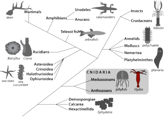
Hydra belongs to Cnidaria, a sister group to bilaterians (Fig.1). The anatomy of Hydra is rather simple: this is basically a bilayered tube that shows an apical-basal polarity, with at one extremity a head region that includes a mouth/anus opening and a ring of tentacles to actively catch the food (Hydra are carnivorous), and at the other extremity a basal disc. Its cellular organization is rather simple, with two cell layers, the ectoderm and the endoderm separated by an extracellular collagenoeous matrix, the mesogolea. Hydra differentiates all cell types required for neuro-muscular transmission, digestion, secretion and sexual reproduction. These different cell types differentiate from three distinct stem cell populations: myoepithelial cells located in the ectoderm, myoepithelial cells located in the endoderm and interstitial cells that are multipotent stem cells giving rise to neurons, mechano-sensory cells (nematocytes or cnidocytes), gland cells and gametes [3, 4].

Over the past 25 years, the genes encoding the signaling proteins that control and execute various cellular behaviors and developmental processes were shown to be present in all animals. The recent sequencing of the genome of two cnidarian species (Nematostella vectensis – sea anemone - and Hydra magnipapillata) indeed confirmed this high level of gene conservation from cnidarians to vertebrates. This definitively supports the relevance of Hydra as a model system to investigate complex biological questions [5, 6].

3. Functional analysis of the genetic circuitry supporting regeneration in Hydra

Gene silencing through RNAi by feeding identifies cellular phenotypes that are shared between Hydra and man

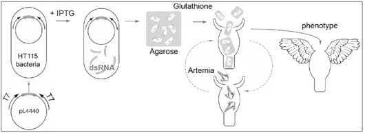
RNAi gene silencing obtained by feeding the animal on dsRNA-producing bacteria was first described in C. elegans and more recently adapted to planarians [7-9]. We adapted this strategy to Hydra (Fig. 2) and proved that this method that is harmless, stepwise and efficient, can induce gene-specific phenotypes [10-13]. In case of the protease inhibitor Kazal1that isspecifically expressed in gland cells of the gastrodermis, RNAi silencing induces a massive autophagy that extends to the digestive cells. This phenotype, which mimics the human SPINK1/mouse SPINK3 pancreatic phenotype, provides the first example of a conserved cellular mechanism from cnidarians to mammals, an example that definitely strengthen the paradigmatic value of this little animal.

This strategy offers the possibility of systematic RNAi screens in Hydra.


Gene silencing by RNA interference in Hydra

Figure 2: Gene silencing by RNA interference in Hydra. The dsRNA feeding strategy developed in nematodes [7, 8] and planarians [9] was adapted to Hydra polyps. Double-stranded RNAs are produced in bacteria and Hydra polyps are repeatedly fed with the bacteria-agarose mixture {Chera, 2006 #212; Buzgariu, 2008 #174}.


4. The apoptosis-dependent activation of the Wnt pathway in head-regenerating tips.

The canonical Wnt pathway is perfectly conserved in Hydra and required for the head to regenerate properly [14, 15]. We recently showed that an asymmetrical wave of apoptosis occurs immediately after mid-gastric bisection affecting 50% of the cells in head-regenerating tips but less than 7% in foot-regenerating ones [13]. Apoptotic cells actually transiently release the Wnt3 signal that activates b-catenin in the surrounding S-phase cells. Indeed progenitors migrate towards the wound, accumulate underneath the apoptotic zone and rapidly divide forming a proliferative zone. Upon inhibition of apoptosis by caspase inhibitors, or upon Wnt3 or b-catenin RNAi silencing, cell proliferation and head regeneration are abolished, whereas simply adding exogenous Wnt3 fully rescues these processes. Conversely
induction of apoptosis in foot-regenerating tips converts them to regenerate heads through the activation of the Wnt3/b-catenin pathway. Hence the level of apoptosis appears critical to trigger compensatory proliferation and head regeneration through the Wnt pathway.


Bi-headed Hydra resulting from the ectopic apoptosis-induced activation of the Wnt3-bcatenin pathway

Figure 3: Bi-headed Hydra resulting from the ectopic apoptosis-induced activation of the Wnt3-bcatenin pathway. Hydra polyps bisected at mid-gastric position regenerate a head from the lower half and a basal disc from the upper one. Here Hydra upper halves were forced to regenerate a head instead of a basal disc. This was obtained by inducing a high level of apoptosis in foot-regenerating tips by briefly heating them immediately after bisection. Ectopic apoptosis leads to the activation of the Wnt3-bcatenin pathway as observed in head-regenerating stumps and therefore converted foot regeneration to head regeneration (see in [1, 2]).


The immediate asymmetric regulation of the MAPK ➜ RSK ➜ CREB ➜ CBP pathway

The MAPK/CREB pathway seems to play a key role in the induction of the head regeneration process. Indeed the cAMP Response Element Binding (CREB) protein is a transcription factor that likely interacts with distinct partners in CRE-binding complexes immediately after bisection [16], and exhibits a series of immediate regulations in the head- but not in the foot-regenerating tips as an RSK-dependent phosphorylation [17] and a rapid up-regulation of the CREB gene expression [18]. In vertebrates, the phosphorylated form of CREB binds to the chromatin modifyer CBP in order to modulate gene expression. Similarly the Hydra CBP encodes a CREB-binding domain and silencing of either RSK or CREB or CBP prevents the immediate wave of apoptosis and the cellular reorganization normally observed in head-regenerating tips (Chera and Galliot, submitted). We are currently investigating how this signaling pathway can sense the stress of amputation to activate a head regeneration program.

The emergence of neurogenesis and apical patterning in metazoans

In Hydra neurogenesis takes place continuously in the adult polyp but the dense apical nervous system also forms de novo in budding and regenerating animals [19- 21]. In bilaterian animals whose ancestor already had a centralized nervous system and sensory organs, the homeobox genes of the ANTP and PAIRED class play a major role in the developing central nervous system [22-24]. Each class includes a large number of families that for most of them diversified quite early in animal evolution, preceding the divergence of Cnidaria [25-27]. This raises the question of the function of these gene families in cnidarians (that differentiate a sophisticated nervous system) and in poriferans (that have no nervous system)[21].

The PAIRED-class homeobox genes

Among evolutionary conserved gene families involved in the specification of brain and sensory organs in bilaterians, several are expressed in the nervous system of cnidarians [28-32]. Among these, the paired-class homeobox gene prdl-a, is expressed in apical nerve cells in adult polyps but after bisection, transiently in endodermal cells of the head-regenerating tip. This result suggested that prdl-a is involved in Hydra head organizer activity through inductive interactions from endoderm to the overlying ectoderm [19, 28]. This result was puzzling as some paired- like genes perform similar tasks at the time head organizer activity is established in mammals, suggesting that molecular mechanisms of anterior patterning can be traced back to cnidarians [33].

The Hox/ParaHox genes

Most ANTP-class gene families do have cnidarian counterparts although Hox-related families are not all present and less similar. In fact Hox-like genes were not found in poriferan, therefore, cnidarian Hox-like genes can be considered as representative of the proto-Hox genes [26, 34-36]. Out of them, cnox-2, theortholog of the paraHox Gsx gene, stimulates interest as its expression is restricted to the precursors of the neuronal and nematocyte cell lineages and cnox-2 is also upregulated during de novo apical neurogenesis at the time head is forming, during budding and in regenerating polyps [11, 26, 37]. When cnox-2 expression is silenced via RNAi, alterations of the apical nervous system and apical patterning process are observed. In vertebrates and Drosophila, Gsx is also involved in neurogenesis [38]. These data suggest an evolutionarily-conserved function for cnox-2/Gsx in neurogenesis and a possible functional link between neurogenesis and apical patterning established quite early in animal evolution [11, 20, 36].

5. References

  1. Galliot, B., and Chera, S. (2010) The Hydra model: disclosing an apoptosis-driven generator of Wnt-based regeneration. Trends Cell Biol in press
  2. Galliot, B., and Ghila, L. (2010) Cell plasticity in homeostasis and regeneration. Mol Reprod Dev in press
  3. Steele, R.E. (2002) Developmental signaling in Hydra: what does it take to build a “simple” animal? Dev Biol 248, 199-219
  4. Galliot, B., Miljkovic-Licina, M., de Rosa, R., and Chera, S. (2006) Hydra, a niche for cell and developmental plasticity. Semin Cell Dev Biol 17, 492-502
  5. Putnam, N.H., Srivastava, M., Hellsten, U., Dirks, B., Chapman, J., Salamov, A., Terry, A., Shapiro, H., et al. (2007) Sea anemone genome reveals ancestral eumetazoan gene repertoire and genomic organization. Science 317, 86-94
  6. Chapman, J.A., Kirkness, E.F., Simakov, O., Hampson, S.E., Mitros, T., Weinmaier, T., Rattei, T., Balasubramanian, P.G., et al. (2010) The dynamic genome of Hydra. Nature 464, 592-596
  7. Timmons, L., Court, D.L., and Fire, A. (2001) Ingestion of bacterially expressed dsRNAs can produce specific and potent genetic interference in Caenorhabditis elegans. Gene 263, 103-112
  8. Kamath, R.S., Martinez-Campos, M., Zipperlen, P., Fraser, A.G., and Ahringer, J. (2001) Effectiveness of specific RNA-mediated interference through ingested double-stranded RNA in Caenorhabditis elegans. Genome Biol 2, RESEARCH0002
  9. Newmark, P.A., Reddien, P.W., Cebria, F., and Sanchez Alvarado, A. (2003) Ingestion of bacterially expressed double-stranded RNA inhibits gene expression in planarians. Proc Natl Acad Sci U S A 100 Suppl 1, 11861-11865
  10. Chera, S., de Rosa, R., Miljkovic-Licina, M., Dobretz, K., Ghila, L., Kaloulis, K., and Galliot, B. (2006)
    Silencing of the hydra serine protease inhibitor Kazal1 gene mimics the human SPINK1 pancreatic phenotype. J Cell Sci 119, 846-857
  11. Miljkovic-Licina, M., Chera, S., Ghila, L., and Galliot, B. (2007) Head regeneration in wild-type hydra requires de novo neurogenesis. Development 134, 1191-1201
  12. Buzgariu, W., Chera, S., and Galliot, B. (2008) Methods to investigate autophagy during starvation and regeneration in hydra. Methods Enzymol 451, 409-437
  13. Chera, S., Ghila, L., Dobretz, K., Wenger, Y., Bauer, C., Buzgariu, W., Martinou, J.C., and Galliot, B. (2009) Apoptotic cells provide an unexpected source of Wnt3 signaling to drive hydra head regeneration. Dev Cell 17, 279-289
  14. Hobmayer, B., Rentzsch, F., Kuhn, K., Happel, C.M., von Laue, C.C., Snyder, P., Rothbacher, U., and Holstein, T.W. (2000) WNT signalling molecules act in axis formation in the diploblastic metazoan Hydra. Nature 407, 186-189
  15. Lengfeld, T., Watanabe, H., Simakov, O., Lindgens, D., Gee, L., Law, L., Schmidt, H.A., Ozbek, S., et al. (2009) Multiple Wnts are involved in Hydra organizer formation and regeneration. Dev Biol 330, 186-199
  16. Galliot, B., Welschof, M., Schuckert, O., Hoffmeister, S., and Schaller, H.C. (1995) The cAMP response element binding protein is involved in hydra regeneration. Development 121, 1205-1216
  17. Kaloulis, K., Chera, S., Hassel, M., Gauchat, D., and Galliot, B. (2004) Reactivation of developmental programs: the cAMP-response element-binding protein pathway is involved in hydra head regeneration. Proc Natl Acad Sci U S A 101, 2363-2368
  18. Chera, S., Kaloulis, K., and Galliot, B. (2007) The cAMP response element binding protein (CREB) as an integrative HUB selector in metazoans: clues from the hydra model system. Biosystems 87, 191-203
  19. Miljkovic-Licina, M., Gauchat, D., and Galliot, B. (2004) Neuronal evolution: analysis of regulatory genes in a first-evolved nervous system, the hydra nervous system. Biosystems 76, 75-87
  20. Galliot, B., Quiquand, M., Ghila, L., de Rosa, R., Miljkovic-Licina, M., and Chera, S. (2009) Origins of neurogenesis, a cnidarian view. Dev Biol 332, 2-24
  21. Galliot, B. (2010) A Key Innovation in Evolution, the Emergence of Neurogenesis: Cellular and Molecular Cues from Cnidarian Nervous Systems. In Key Transitions in Animal Evolution (Schierwater, B., and De Salle, R., eds), 127-161, Science Publishers & CRC Press
  22. Pichaud, F., and Desplan, C. (2002) Pax genes and eye organogenesis. Curr Opin Genet Dev 12, 430-434
  23. Hirth, F., Kammermeier, L., Frei, E., Walldorf, U., Noll, M., and Reichert, H. (2003) An urbilaterian origin of the tripartite brain: developmental genetic insights from Drosophila. Development 130, 2365-2373
  24. Denes, A.S., Jekely, G., Steinmetz, P.R., Raible, F., Snyman, H., Prud'homme, B., Ferrier, D.E., Balavoine, G., et al. (2007) Molecular architecture of annelid nerve cord supports common origin of nervous system centralization in bilateria. Cell 129, 277-288
  25. Galliot, B., de Vargas, C., and Miller, D. (1999) Evolution of homeobox genes: Q50 Paired-like genes founded the Paired class. Dev Genes Evol 209, 186-197
  26. Gauchat, D., Mazet, F., Berney, C., Schummer, M., Kreger, S., Pawlowski, J., and Galliot, B. (2000)
    Evolution of Antp-class genes and differential expression of Hydra Hox/paraHox genes in anterior patterning. Proc Natl Acad Sci U S A 97, 4493-4498
  27. Ryan, J.F., Burton, P.M., Mazza, M.E., Kwong, G.K., Mullikin, J.C., and Finnerty, J.R. (2006) The cnidarian-bilaterian ancestor possessed at least 56 homeoboxes. Evidence from the starlet sea anemone, Nematostella vectensis. Genome Biol 7, R64
  28. Gauchat, D., Kreger, S., Holstein, T., and Galliot, B. (1998) prdl-a, a gene marker for hydra apical differentiation related to triploblastic paired-like head-specific genes. Development 125, 1637-1645
  29. Lindgens, D., Holstein, T.W., and Technau, U. (2004) Hyzic, the Hydra homolog of the zic/odd- paired gene, is involved in the early specification of the sensory nematocytes. Development 131, 191-201
  30. Gauchat, D., Escriva, H., Miljkovic-Licina, M., Chera, S., Langlois, M.C., Begue, A., Laudet, V., and Galliot, B. (2004) The orphan COUP-TF nuclear receptors are markers for neurogenesis from cnidarians to vertebrates. Dev Biol 275, 104-123
  31. Stierwald, M., Yanze, N., Bamert, R.P., Kammermeier, L., and Schmid, V. (2004) The Sine oculis/Six class family of homeobox genes in jellyfish with and without eyes: development and eye regeneration. Dev Biol 274, 70-81
  32. Marlow, H.Q., Srivastava, M., Matus, D.Q., Rokhsar, D., and Martindale, M.Q. (2009) Anatomy and development of the nervous system of Nematostella vectensis, an anthozoan cnidarian. Dev Neurobiol 69, 235-254
  33. Galliot, B., and Miller, D. (2000) Origin of anterior patterning. How old is our head? Trends Genet 16, 1-5
  34. Chourrout, D., Delsuc, F., Chourrout, P., Edvardsen, R.B., Rentzsch, F., Renfer, E., Jensen, M.F., Zhu, B., et al. (2006) Minimal ProtoHox cluster inferred from bilaterian and cnidarian Hox complements. Nature 442, 684-687
  35. Chiori, R., Jager, M., Denker, E., Wincker, P., Da Silva, C., Le Guyader, H., Manuel, M., and Queinnec, E. (2009) Are Hox genes ancestrally involved in axial patterning? Evidence from the hydrozoan Clytia hemisphaerica (Cnidaria). PLoS ONE 4, e4231
  36. Quiquand, M., Yanze, N., Schmich, J., Schmid, V., Galliot, B., and Piraino, S. (2009) More constraint on ParaHox than Hox gene families in early metazoan evolution. Dev Biol 328, 173-187
  37. Schummer, M., Scheurlen, I., Schaller, C., and Galliot, B. (1992) HOM/HOX homeobox genes are present in hydra (Chlorohydra viridissima) and are differentially expressed during regeneration. Embo J 11, 1815-1823
  38. Mieko Mizutani, C., and Bier, E. (2008) EvoDevo: the origins of BMP signalling in the neuroectoderm. Nat Rev Genet 9, 663-677

Team

Brigitte Galliot
Emeritus Professor Leader

Prof Brigitte Galliot

Publications

Page
Cells & development
Authors: Galliot, Wenger
View Publication
The experimental and conceptual knowledge in 1909 led to the discovery of the Hydra head organizer through transplantation experiments between pigmented and non-pigmented animals; a discovery followed by numerous transplantations demonstrating cross-regulation between activating and inhibiting components distributed along the body axis. This experimental work inspired mathematicians, engineers, physicists and computer scientists to develop theoretical models predicting the principles of developmental mechanisms. Today, we know that the Wnt/β-catenin/Sp5/Zic4 gene regulatory network (GRN) links organizer activity, morphogenesis and cellular identity in Hydra, with variable conformations depending on the region or epithelial layer, and varied phenotypes depending on which GRN element is misregulated. In intact animals, Wnt/β-catenin signaling acts as the head activator at the tip of the hypostome, restricted by Sp5 in the other regions of the animal. Moreover, in the tentacle ring, Sp5 and Zic4 act epistatically to support tentacle differentiation and prevent basal disc differentiation. Along the body column, Sp5 is self-repressed in the epidermis and acts as a head inhibitor along the gastrodermis. Other players modulate these activities, such as TSP and Margin/RAX apically, Notch signaling in the tentacle zone, Dkk1/2/4 and HAS-7 in the body column. In the developmental context of regeneration, cells below the amputation zone switch from repressed to locally de novo activated head organizer status, a transition driven by immediate symmetrical and asymmetrical metabolic changes that lead to gene expression regulations involving components and modulators of Wnt/β-catenin signaling, early-pulse and early-late transient both often symmetrical, together with sustained ones, specific to head regeneration.
Biomedicines
Authors: Iglesias Ollé L, Perruchoud C, Sanchez PGL, Vogg MC, Galliot B
View Publication
head formation depends on an organizing center in which Wnt/β-catenin signaling, that plays an inductive role, positively regulates and , with Sp5 limiting expression and Zic4 triggering tentacle formation. Using transgenic lines in which the promoter drives eGFP expression in either the epidermis or gastrodermis, we show that promoter activity is differentially regulated in each epithelial layer. In intact animals, epidermal GFP activity is strong apically and weak along the body column, while in the gastrodermis, it is maximal in the tentacle ring region and maintained at a high level along the upper body column. During apical regeneration, :GFP is activated early in the gastrodermis and later in the epidermis. Alsterpaullone treatment induces a shift in apical expression towards the body column where it forms transient circular figures in the epidermis. Upon (RNAi), GFP activity is down-regulated in the epidermis while bud-like structures expressing GFP in the gastrodermis develop. (RNAi) reveals a negative autoregulation in the epidermis, but not in the gastrodermis. These differential regulations in the epidermis and gastrodermis highlight the distinct architectures of the network in the hypostome, tentacle base and body column of intact animals, as well as in the buds and apical and basal regenerating tips.
Science advances
Authors: Vogg MC, Ferenc J, Buzgariu WC, Perruchoud C, Sanchez PGL, Beccari L, Nuninger C, Le Cras Y, Delucinge-Vivier C, Papasaikas P, Vincent S, Galliot B, Tsiairis CD
View Publication
The molecular mechanisms that maintain cellular identities and prevent dedifferentiation or transdifferentiation remain mysterious. However, both processes are transiently used during animal regeneration. Therefore, organisms that regenerate their organs, appendages, or even their whole body offer a fruitful paradigm to investigate the regulation of cell fate stability. Here, we used as a model system and show that Zic4, whose expression is controlled by Wnt3/β-catenin signaling and the Sp5 transcription factor, plays a key role in tentacle formation and tentacle maintenance. Reducing expression suffices to induce transdifferentiation of tentacle epithelial cells into foot epithelial cells. This switch requires the reentry of tentacle battery cells into the cell cycle without cell division and is accompanied by degeneration of nematocytes embedded in these cells. These results indicate that maintenance of cell fate by a Wnt-controlled mechanism is a key process both during homeostasis and during regeneration.
Methods in molecular biology (Clifton, N.J.)
Authors: Vogg MC, Galliot B
View Publication
In addition to its ability to regenerate any amputated body part, the Hydra freshwater polyp shows the amazing ability to regenerate as a full polyp after a complete dissociation of its tissues. The developmental processes at work in reaggregates undergoing whole-body regeneration can be investigated at the molecular level by RNA interference (RNAi). Here we provide a protocol that combines β-catenin RNAi with reaggregation. This protocol serves as a basis to generate "RNAi-reaggregates," followed by the extraction of high-quality RNA for the precise quantification of gene expression by real-time PCR. This protocol is efficient, providing both a molecular signature, with the significant downregulation of β-catenin and Wnt3, as well as a robust phenotype, the lack of axis formation, which is observed in all reaggregates.
Methods in molecular biology (Clifton, N.J.)
Authors: Buzgariu W, Aubry-Lachainaye JP, Galliot B
View Publication
The freshwater Hydra polyp is a versatile model to study whole-body regeneration from a developmental as well as a cellular point of view. The outstanding regenerative capacities of Hydra are based on its three populations of adult stem cells located in the central body column of the animal. There, these three populations, gastrodermal epithelial, epidermal epithelial, and interstitial, continuously cycle in homeostatic conditions, and their activity is locally regulated after mid-gastric bisection. Moreover, they present an unusual cycling behavior with a short G1 phase and a pausing in G2. This particular cell cycle has been studied for a long time with classical microscopic methods. We describe here two flow cytometry methods that provide accurate and reproducible quantitative data to monitor cell cycle regulation in homeostatic and regenerative contexts. We also present a cell sorting procedure based on flow cytometry, whereby stem cells expressing a fluorescent reporter protein in transgenic lines can be enriched for use in applications such as transcriptomic, proteomic, or cell cycle analysis.
Cold Spring Harbor perspectives in biology
Authors: Vogg MC, Buzgariu W, Suknovic NS, Galliot B
View Publication
Here we discuss the developmental and homeostatic conditions necessary for regeneration. is characterized by populations of adult stem cells paused in the G2 phase of the cell cycle, ready to respond to injury signals. The body column can be compared to a blastema-like structure, populated with multifunctional epithelial stem cells that show low sensitivity to proapoptotic signals, and high inducibility of autophagy that promotes resistance to stress and starvation. Intact polyps also exhibit a dynamic patterning along the oral-aboral axis under the control of homeostatic organizers whose activity results from regulatory loops between activators and inhibitors. As in bilaterians, injury triggers the immediate production of reactive oxygen species (ROS) signals that promote wound healing and contribute to the reactivation of developmental programs via cell death and the de novo formation of new organizing centers from somatic tissues. In aging , regeneration is rapidly lost as homeostatic conditions are no longer pro-regenerative.
Mechanisms of ageing and development
Authors: Suknovic N, Tomczyk S, Colevret D, Perruchoud C, Galliot B
View Publication
Hydra vulgaris (Hv) has a high regenerative potential and negligible senescence, as its stem cell populations divide continuously. In contrast, the cold-sensitive H. oligactis (Ho_CS) rapidly develop an aging phenotype under stress, with epithelial stem cells deficient for autophagy, unable to maintain their self-renewal. Here we tested in aging, non-aging and regenerating Hydra the activity and regulation of the ULK1 kinase involved in autophagosome formation. In vitro kinase assays show that human ULK1 activity is activated by Hv extracts but repressed by Ho_CS extracts, reflecting the ability or inability of their respective epithelial cells to initiate autophagosome formation. The factors that keep ULK1 inactive in Ho_CS remain uncharacterized. Hv_Basel1 animals exposed to the ULK1 inhibitor SBI-0206965 no longer regenerate their head, indicating that the sustained autophagy flux recorded in regenerating Hv_AEP2 transgenic animals expressing the DsRed-GFP-LC3A autophagy tandem sensor is necessary. The SBI-0206965 treatment also alters the contractility of intact Hv_Basel1 animals, and leads to a progressive reduction of animal size in Hv_AEP2, similarly to what is observed in ULK1(RNAi) animals. We conclude that the evolutionarily-conserved role of ULK1 in autophagy initiation is crucial to maintain a dynamic homeostasis in Hydra, which supports regeneration efficiency and prevents aging.
PloS one
Authors: Schenkelaars Q, Perez-Cortes D, Perruchoud C, Galliot B
View Publication
Hydra are freshwater polyps widely studied for their amazing regenerative capacity, adult stem cell populations, low senescence and value as ecotoxicological marker. Many wild-type strains of H. vulgaris have been collected worldwide and maintained effectively under laboratory conditions by asexual reproduction, while stable transgenic lines have been continuously produced since 2006. Efforts are now needed to ensure the genetic characterization of all these strains, which despite similar morphologies, show significant variability in their response to gene expression silencing procedures, pharmacological treatments or environmental conditions. Here, we established a rapid and reliable procedure at the single polyp level to produce via PCR amplification of three distinct microsatellite sequences molecular signatures that distinguish between Hydra strains and species. The TG-rich region of an uncharacterized gene (ms-c25145) helps to distinguish between Eurasian H. vulgaris-Pallas strains (Hm-105, Basel1, Basel2 and reg-16), between Eurasian and North American H. vulgaris strains (H. carnea, AEP), and between the H. vulgaris and H. oligactis species. The AT-rich microsatellite sequences located in the AIP gene (Aryl Hydrocarbon Receptor Interaction Protein, ms-AIP) also differ between Eurasian and North American H. vulgaris strains. Finally, the AT-rich microsatellite located in the Myb-Like cyclin D-binding transcription factor1 gene (ms-DMTF1) gene helps to distinguish certain transgenic AEP lines. This study shows that the analysis of microsatellite sequences, which is capable of tracing genomic variations between closely related lineages of Hydra, provides a sensitive and robust tool for characterizing the Hydra strains.
Development (Cambridge, England)
Authors: Tomczyk S, Suknovic N, Schenkelaars Q, Wenger Y, Ekundayo K, Buzgariu W, Bauer C, Fischer K, Austad S, Galliot B
View Publication
possesses three distinct stem cell populations that continuously self-renew and prevent aging in However sexual animals from the cold-sensitive ( strain develop an aging phenotype upon gametogenesis induction, initiated by the loss of interstitial stem cells. Animals stop regenerating, lose their active behaviors and die within three months. This phenotype is not observed in the cold-resistant strain. To dissect the mechanisms of aging we compared the self-renewal of epithelial stem cells in these two strains and found it irreversibly reduced in aging while sustained in non-aging We also identified a deficient autophagy in epithelial cells, with a constitutive deficiency in autophagosome formation as detected with the mCherry-eGFP-LC3A/B autophagy sensor, an inefficient response to starvation as evidenced by the accumulation of the autophagosome cargo protein p62/SQSTM1, and a poorly-inducible autophagy flux upon proteasome inhibition. In the non-aging animals, the blockade of autophagy by knocking-down suffices to induce aging. This study highlights the essential role of a dynamic autophagy flux to maintain epithelial stem cell renewal and prevent aging.
Development (Cambridge, England)
Authors: Vogg MC, Galliot B, Tsiairis CD
View Publication
The freshwater polyp provides a potent model system for investigating the conditions that promote wound healing, reactivation of a developmental process and, ultimately, regeneration of an amputated body part. polyps can also be dissociated to the single cell level and can regenerate a complete body axis from aggregates, behaving as natural organoids. In recent years, the ability to exploit has been expanded with the advent of new live-imaging approaches, genetic manipulations that include stable transgenesis, gene silencing and genome editing, and the accumulation of high-throughput omics data. In this Primer, we provide an overview of as a model system for studying regeneration, highlighting recent results that question the classical self-enhancement and long-range inhibition model supposed to drive regeneration. We underscore the need for integrative explanations incorporating biochemical as well as mechanical signalling.
Methods in molecular biology (Clifton, N.J.)
Authors: Buzgariu W, Curchod ML, Perruchoud C, Galliot B
View Publication
The nervous system is produced and maintained in adult Hydra through the continuous production of nerve cells and mechanosensory cells (nematocytes or cnidocytes). De novo neurogenesis occurs slowly in intact animals that replace their dying nerve cells, at a faster rate in animals regenerating their head as a complete apical nervous system is built in few days. To dissect the molecular mechanisms that underlie these properties, a precise monitoring of the markers of neurogenesis and nematogenesis is required. Here we describe the conditions for an efficient BrdU-labeling coupled to an immunodetection of neuronal markers, either regulators of neurogenesis, here the homeoprotein prdl-a, or neuropeptides such as RFamide or Hym-355. This method can be performed on whole-mount animals as well as on macerated tissues when cells retain their morphology. Moreover, when antibodies are not available, BrdU-labeling can be combined with the analysis of gene expression by whole-mount in situ hybridization. This co-immunodetection procedure is well adapted to visualize and quantify the dynamics of de novo neurogenesis. Upon continuous BrdU labeling, the repeated measurements of BrdU-labeling indexes in specific cellular populations provide a precise monitoring of nematogenesis as well as neurogenesis, in homeostatic or developmental conditions.
Developmental neurobiology
Authors: Tomczyk S, Buzgariu W, Perruchoud C, Fisher K, Austad S, Galliot B
View Publication
In Hydra the nervous system is composed of neurons and mechanosensory cells that differentiate from interstitial stem cells (ISCs), which also provide gland cells and germ cells. The adult nervous system is actively maintained through continuous de novo neurogenesis that occurs at two distinct paces, slow in intact animals and fast in regenerating ones. Surprisingly Hydra vulgaris survive the elimination of cycling interstitial cells and the subsequent loss of neurogenesis if force-fed. By contrast, H. oligactis animals exposed to cold temperature undergo gametogenesis and a concomitant progressive loss of neurogenesis. In the cold-sensitive strain Ho_CS, this loss irreversibly leads to aging and animal death. Within four weeks, Ho_CS animals lose their contractility, feeding response, and reaction to light. Meanwhile, two positive regulators of neurogenesis, the homeoprotein prdl-a and the neuropeptide Hym-355, are no longer expressed, while the "old" RFamide-expressing neurons persist. A comparative transcriptomic analysis performed in cold-sensitive and cold-resistant strains confirms the downregulation of classical neuronal markers during aging but also shows the upregulation of putative regulators of neurotransmission and neurogenesis such as AHR, FGFR, FoxJ3, Fral2, Jagged, Meis1, Notch, Otx1, and TCF15. The switch of Fral2 expression from neurons to germ cells suggests that in aging animals, the neurogenic program active in ISCs is re-routed to germ cells, preventing de novo neurogenesis and impacting animal survival.
Nature communications
Authors: Vogg MC, Beccari L, Iglesias Ollé L, Rampon C, Vriz S, Perruchoud C, Wenger Y, Galliot B
View Publication
Polyps of the cnidarian Hydra maintain their adult anatomy through two developmental organizers, the head organizer located apically and the foot organizer basally. The head organizer is made of two antagonistic cross-reacting components, an activator, driving apical differentiation and an inhibitor, preventing ectopic head formation. Here we characterize the head inhibitor by comparing planarian genes down-regulated when β-catenin is silenced to Hydra genes displaying a graded apical-to-basal expression and an up-regulation during head regeneration. We identify Sp5 as a transcription factor that fulfills the head inhibitor properties: leading to a robust multiheaded phenotype when knocked-down in Hydra, acting as a transcriptional repressor of Wnt3 and positively regulated by Wnt/β-catenin signaling. Hydra and zebrafish Sp5 repress Wnt3 promoter activity while Hydra Sp5 also activates its own expression, likely via β-catenin/TCF interaction. This work identifies Sp5 as a potent feedback loop inhibitor of Wnt/β-catenin signaling, a function conserved across eumetazoan evolution.
Current opinion in neurobiology
Authors: Rentzsch F, Juliano C, Galliot B
View Publication
Cnidarians shared a common ancestor with bilaterians more than 600 million years ago. This sister group relationship gives them an informative phylogenetic position for understanding the fascinating morphological and molecular cell type diversity of bilaterian nervous systems. Moreover, cnidarians display novel features such as endodermal neurogenesis and independently evolved centralizations, which provide a platform for understanding the evolution of nervous system innovations. In recent years, the application of modern genomic tools has significantly advanced our understanding of cnidarian nervous system structure and function. For example, transgenic reporter lines and gene knockdown experiments in several cnidarian species reveal a significant degree of conservation in the neurogenesis gene regulatory program, while single cell RNA sequencing projects are providing a much deeper understanding of cnidarian neural cell type diversity. At the level of neural function, the physiological properties of ion channels have been described and calcium imaging of the nervous system in whole animals has allowed for the identification of neural circuits underlying specific behaviours. Cnidarians have arrived in the modern era of molecular neurobiology and are primed to provide exciting new insights into the early evolution of nervous systems.
The International journal of developmental biology
Authors: Galliot B, Buzgariu W, Schenkelaars Q, Wenger Y
View Publication
An essential dimension of 3D regeneration in adult animals is developmental, with the formation of organizers from somatic tissues. These organizers produce signals that recruit surrounding cells and drive the restoration of the missing structures (organs, appendages, body parts). However, even in animals with a high regenerative potential, this developmental potential is not sufficient to achieve regeneration as homeostatic conditions at the time of injury need to be "pro-regenerative". In Hydra, we identified four distinct homeostatic properties that provide a pro-regenerative framework and we discuss here how these non-developmental properties impact regeneration. First, both the epithelial and the interstitial-derived cells are highly plastic along the animal body, a plasticity that offers several routes to achieve regeneration. Second, the abundant stocks of continuously self-renewing adult stem cells form a constitutive pro-blastema in the central body column, readily activated upon bisection. Third, the autophagy machinery in epithelial cells guarantees a high level of fitness and adaptation to detrimental environmental conditions, as evidenced by the loss of regeneration in animals where autophagy is dysfunctional. Fourth, the extracellular matrix, named mesoglea in Hydra, provides a dynamically-patterned environment where the molecular and mechanical signals induced by injury get translated into a regenerative process. We claim that these homeostatic pro-regenerative features contribute to define the high regenerative potential of adult Hydra.
Conn’s Handbook for models on human aging (Second Edition)
Authors: Schenkelaars Q., Tomczyk S., Wenger Y., Ekundayo K., Girard V., Buzgariu W., Austad S. and Galliot B.
View Publication
Conn's Handbook of Models for Human Aging, Second Edition, presents key aspects of biology, nutrition, factors affecting lifespan, methods of age determination, use in research and the disadvantages/advantages of use. Using a multidisciplinary approach, this updated edition is designed as the only comprehensive, current work that covers the diversity in aging models. Chapters on comparative models explore age-related diseases, including Alzheimer's, joint disease, cataracts, cancer and obesity. Also included are new tricks and approaches not available in primary publications. This must-have handbook is an indispensable resource for researchers interested in the mechanisms of aging, gerontologists, health professionals, allied health practitioners and students.
Developmental biology
Authors: Buzgariu W, Wenger Y, Tcaciuc N, Catunda-Lemos AP, Galliot B
View Publication
Hydra tissues are made from three distinct populations of stem cells that continuously cycle and pause in G2 instead of G1. To characterize the role of cell proliferation after mid-gastric bisection, we have (i) used flow cytometry and classical markers to monitor cell cycle modulations, (ii) quantified the transcriptomic regulations of 202 genes associated with cell proliferation during head and foot regeneration, and (iii) compared the impact of anti-proliferative treatments on regeneration efficiency. We confirm two previously reported events: an early mitotic wave in head-regenerating tips, when few cell cycle genes are up-regulated, and an early-late wave of proliferation on the second day, preceded by the up-regulation of 17 cell cycle genes. These regulations appear more intense after mid-gastric bisection than after decapitation, suggesting a position-dependent regulation of cell proliferation during head regeneration. Hydroxyurea, which blocks S-phase progression, delays head regeneration when applied before but not after bisection. This result is consistent with the fact that the Hydra central region is enriched in G2-paused adult stem cells, poised to divide upon injury, thus forming a necessary constitutive pro-blastema. However a prolonged exposure to hydroxyurea does not block regeneration as cells can differentiate apical structures without traversing S-phase, and also escape in few days the hydroxyurea-induced S-phase blockade. Thus Hydra head regeneration, which is a fast event, is highly plastic, relying on large stocks of adult stem cells paused in G2 at amputation time, which immediately divide to proliferate and/or differentiate apical structures even when S-phase is blocked.
Development (Cambridge, England)
Authors: Galliot B, Crescenzi M, Jacinto A, Tajbakhsh S
View Publication
The 6th EMBO conference on the Molecular and Cellular Basis of Regeneration and Tissue Repair took place in Paestum (Italy) on the 17th-21st September, 2016. The 160 scientists who attended discussed the importance of cellular and tissue plasticity, biophysical aspects of regeneration, the diverse roles of injury-induced immune responses, strategies to reactivate regeneration in mammals, links between regeneration and ageing, and the impact of non-mammalian models on regenerative medicine.
Current topics in developmental biology
Authors: Vogg MC, Wenger Y, Galliot B
View Publication
The growth and patterning of anatomical structures from specific cellular fields in developing organisms relies on organizing centers that instruct surrounding cells to modify their behavior, namely migration, proliferation, and differentiation. We discuss here how organizers can form in adult organisms, a process of utmost interest for regenerative medicine. Animals like Hydra and planarians, which maintain their shape and fitness thanks to a highly dynamic homeostasis, offer a useful paradigm to study adult organizers in steady-state conditions. Beside the homeostatic context, these model systems also offer the possibility to study how organizers form de novo from somatic adult tissues. Both extracellular matrix remodeling and caspase activation play a key role in this transition, acting as promoters of organizer formation in the vicinity of the wound. Their respective roles and the crosstalk between them just start to be deciphered.
Tissue barriers
Authors: Buzgariu W, Al Haddad S, Tomczyk S, Wenger Y, Galliot B
View Publication
Epithelial sheets, a synapomorphy of all metazoans but porifers, are present as 2 layers in cnidarians, ectoderm and endoderm, joined at their basal side by an extra-cellular matrix named mesoglea. In the Hydra polyp, epithelial cells of the body column are unipotent stem cells that continuously self-renew and concomitantly express their epitheliomuscular features. These multifunctional contractile cells maintain homeostasis by providing a protective physical barrier, by digesting nutrients, by selecting a stable microbiota, and by rapidly closing wounds. In addition, epithelial cells are highly plastic, supporting the adaptation of Hydra to physiological and environmental changes, such as long starvation periods where survival relies on a highly dynamic autophagy flux. Epithelial cells also play key roles in developmental processes as evidenced by the organizer activity they develop to promote budding and regeneration. We propose here an integrative view of the homeostatic and developmental aspects of epithelial plasticity in Hydra.

More Information

Order hydras

You can order hydras by clicking this link.

Highlights in General Media about our Research (selection)

2017

Radio RTS la 1ère, CQFD by Silvio Dolzan, Pierre Bratschi. Il reste de la vie après la mort. Oct. 30 (9 min). https://www.rts.ch/play/radio/cqfd/audio/il-reste-de-la-vie-apres-la-mort?id=9002141

Le Temps, Brigitte Galliot et Denis Duboule: en constante évolution. by Sylvie Logean, Oct. 3. https://www.letemps.ch/sciences/2017/10/03/brigitte-galliot-denis-duboule-constante-evolution

2016

Radio RTS la 1ère, CQFD Rencontre avec Brigitte Galliot emission de S. Gabioud avec Adrien Zerbini (36 min). http://www.rts.ch/la-1ere/programmes/cqfd/8055408-rencontre-avec-brigitte-galliot-14-10-2016.html

2015 

Online Movie on the AcademiaNet Project (database of excellent female scientists). Bosch Stiftung. http://www.academia-net.org/project/

Jeunesse éternelle. Simples Immortels. Documentary movie by Romain Miranda for TSR (TV Swiss channel), “Mise au Point” Oct. 25, 2015. http://www.rts.ch/emissions/mise-au-point/7086228-conseil-federal-peages-urbains-jeunesse-eternelle.html

Interview on the channel RTS (Radio Télévision Suisse Romande), CQFD program 2015. Journalist: Bastien Confino.
L’hydre modifie son programme génétique

  • http://www.rts.ch/la-1ere/programmes/cqfd/7237869-l-hydre-modifie-son-programme-genetique-23-11-2015.html 
  • interview accessible on the site Avis d’Experts: http://avisdexperts.ch/videos/view/4613/10 (10 minutes)
    • Hydra can modify its genetic program. Science Daily, 23.11.2015
    • Las hidras pueden modificar su programa genético. Noticias de la Ciencia y la Tecnologia, 26.11.2015
    • Immortal Hydra Is Able To Genetically Modify Itself. IFL Science, 26.11.2015
    • Hydra genetically reprograms skin cells after losing its nerve. The Guardian, 27.11.2015
    • Comment survivre sans neurones. Pour la Science, 03.12.2015
    • L’hydre peut vivre sans cerveau. Elle n’est pas la seule. Le Matin, 04.12.2015
    • Ein leuchtender Jungbrunnen.  faz.net / Frankfurter Allgemeine Zeitung Online, 28.11.2015

2012

Interview on the channel RTS (Radio Télévision Suisse Romande), CQFD program 2015. Journalist: Bastien Confino.
L'hydre, du mythe à la science : http://www.rts.ch/la-1ere/programmes/cqfd/4469156-l-hydre-du-mythe-a-la-science-13-12-2012.html

2009

Report on the Evening News at the Swiss TV of the discovery of cell death and regeneration. DÉCOUVERTE SUR LA FACULTÉ DE RÉGÉNÉRATION DE L'HYDRE
This interview is accessible on the site Avis d’Experts: http://www.avisdexperts.ch/videos/view/925  (2’03 minutes)


Movies 

1999     

Galliot B. L’hydre, un embryon immortel.  (8 min) Université de Genève

2003     

Regeneration and Stem Cells (52 min) Documentary movie directed by Jean-Marie Cornuel produced by Télé-Images-Nature. www.teleimages.com/newsletter.htm

2007

L’immortalité? (70 min). 36°9, produced and directed by Mario Fossatti & Isabelle Moncada. Documentary movie produced by the TSR (Télévision Suisse Romande). (French)
http://www.rts.ch/play/tv/36-9/video/le-secret-de-lhydre?id=57661

2008    

Quand la science va à la plage!   Documentary movie directed by Claude-Julie Parisot, produced by KAMI products for ARTE channel.
http://boutique.arte.tv/f2355-quandlasciencevaalaplage

2009   

Einstein: Der Polyp mit dem Ewigen Leben. (3.30 min) Documentary movie produced by SF (Schweizer Fernsehen).

2014     

Immortels. (52 min) Directed by Sarah Lainé. Coproduction : DOCLAND YARD, AB PRODUCTIONS  for Encyclo (Science et Vie TV), a channel dedicated to discovery and scientific knowledge (French)
http://www.gedeonprogrammes.com/wp-content/uploads/2014/01/CATALOGUE-GEDEON_2_VF_BD2.pdf


Publications as Guest Editor of special issues

Galliot, B, Tanaka, E.M. Simon A. guest editors of “Cellular and Molecular Basis of Regeneration and Tissue Repair”. Cellular and Molecular Life Sciencesvol. 65, issue 1 (9 articles).

Galliot B. guest editor of « The Hydra model system » International Journal of Developmental Biology, vol 56, issues 6/7/8 (22 articles).

Galliot B. guest editor of “Mechanisms of Regeneration” (11 articles) Current Topics in Developmental Biology, 108,  http://www.sciencedirect.com/science/bookseries/00702153


Articles on Science and Society

2003

Galliot B. A lire avant de vous lancer dans une carrière de chercheuse. Médecine & Hygiène, 2459, 2303-2306. https://www.revmed.ch/RMS/2003/RMS-2459/1215

2007

Galliot B. La culture scientifique pour une honnête femme du 21ème siècle, est-ce encore possible ? édité par J-J Forney, Fondation Culture & Rencontre, Genève. https://archive-ouverte.unige.ch/unige:38931

2008

Gehring W, Galliot B and Piraino S. In memoriam: Volker Schmid. Int J Dev Biol, 52, 1013 – 1014.http://www.ijdb.ehu.es/web/paper/082708wg/in-memoriam-volker-schmid-1939-2008

2016

Lievens P, Galliot B, Floors F et al. Fundamental Research in HORIZON2020. LERU paper, in press.

Lievens P, Galliot B, Floors F et al. (LERU natural sciences working group). The strength of collaborative research for discovery in HORIZON2020. LERU note, https://www.leru.org/files/The-Strength-of-Collaborative-Research-for-Discovery-in-Horizon-2020-Full-paper.pdf

2017

Väänänen J, van Tienderen P, et al. (LERU Natural Sciences policy group). Four Golden Principles for enhancing the quality, access and impact of research infrastructures LERU note, https://www.leru.org/publications/four-golden-principles-for-enhancing-the-quality-access-and-impact-of-research-infrastructures